Tested applications:WBIHC-PIF/ICCIPChIPChIP-seqRIPFCFC(Intra)ELISAMeDIPNucleotide ArrayDBFACSCoIPCUT&TagmeRIPInhibitionReactivity:Human, Mouse, Rat
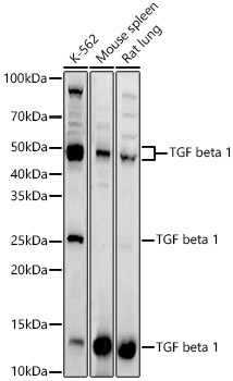
Western blot analysis of various lysates using [KD Validated] TGF beta 1 Rabbit mAb (A25313) at 1:1000 dilution.
Secondary antibody: HRP-conjugated Goat anti-Rabbit IgG (H+L) (AS014) at 1:10000 dilution.
Lysates / proteins: 25 μg per lane.
Blocking buffer: 3 % nonfat dry milk in TBST.
Detection: ECL Basic Kit (RM00020).
Exposure time: 90s.
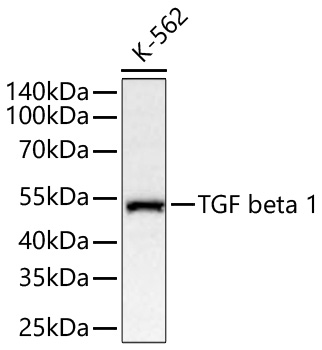
Western blot analysis of lysates from wild type (WT) and TGF beta 1 knockdown (KD) 293T cells using [KD Validated] TGF beta 1 Rabbit mAb (A25313) at 1:1000 dilution incubated overnight at 4℃.
Secondary antibody: HRP-conjugated Goat anti-Rabbit IgG (H+L) (AS014) at 1:10000 dilution.
Lysates/proteins: 25 μg per lane.
Blocking buffer: 3% nonfat dry milk in TBST.
Detection: ECL Basic Kit (RM00020).
Exposure time: 90s.
Immunohistochemistry analysis of paraffin-embedded Human small intestine tissue using [KD Validated] TGF beta 1 Rabbit mAb (A25313) at a dilution of 1:100 (40x lens). High pressure antigen retrieval was performed with 0.01 M Tris-EDTA buffer (pH 9.0) prior to IHC staining.
Immunohistochemistry analysis of paraffin-embedded Mouse spleen tissue using [KD Validated] TGF beta 1 Rabbit mAb (A25313) at a dilution of 1:100 (40x lens). High pressure antigen retrieval was performed with 0.01 M Tris-EDTA buffer (pH 9.0) prior to IHC staining.
| Product name | [KD Validated] TGF beta 1 Rabbit mAb |
|---|---|
| Catalog No. | A25313 |
| Host species | Rabbit |
| Purification method | Affinity purification |
| Isotype | IgG |
| CloneNo. | ARC3248 |
| Immunogen | Recombinant protein |
|---|---|
| Sequence | Email for sequence |
| Gene ID | |
| Swiss Prot | |
| Synonyms | CED; LAP; DPD1; TGFB; IBDIMDE; TGFbeta; TGF-beta1; TGF beta 1 |
| Calculated MW | 44kDa |
| Observed MW | 12kDa/44kDa/12kDa, 44kDa |
| Reactivity | Human, Mouse, Rat |
|---|---|
| Tested applications | WBIHC-PIF/ICCIPChIPChIP-seqRIPFCFC(Intra)ELISAMeDIPNucleotide ArrayDBFACSCoIPCUT&TagmeRIPInhibition |
| Recommended dilution |
|
| Storage buffer | Store at -20℃. Avoid freeze / thaw cycles. Buffer: PBS with 0.09% Sodium azide, 0.05% BSA, 50% glycerol, pH7.3. |
| Key application | Western blotting Immunohistochemistry |
| Positive samples | A549, K-562, Rat spleen, 293T |
| Cellular location | Secreted, extracellular matrix, extracellular space. |
| Customer validation | IHC(Rattus norvegicus) WB(Mus musculus, Homo sapiens) |
To download a Certificate of Compliance, please enter your Lot number below:
Lot number
* For research use only. Not for therapeutic or diagnostic purposes.
Publishing research using A25313? Please let us know so that we can cite the reference in this datasheet.
![ABclonal:Western blot - [KD Validated] TGF beta 1 Rabbit mAb (A25313)](https://img.abclonal.com/abclonal-manage/Catalog/A25313/A25313_1.jpg?t=1739264338 "ABclonal:Western blot - [KD Validated] TGF beta 1 Rabbit mAb (A25313)")
![ABclonal:Western blot - [KD Validated] TGF beta 1 Rabbit mAb (A25313)](https://img.abclonal.com/abclonal-manage/Catalog/A25313/A25313_2.jpg?t=1739264338 "ABclonal:Western blot - [KD Validated] TGF beta 1 Rabbit mAb (A25313)")
![ABclonal:Immunohistochemistry - [KD Validated] TGF beta 1 Rabbit mAb (A25313)](https://img.abclonal.com/abclonal-manage/Catalog/A25313/A25313_3.jpg?t=1739264338 "ABclonal:Immunohistochemistry - [KD Validated] TGF beta 1 Rabbit mAb (A25313)")
![ABclonal:Immunohistochemistry - [KD Validated] TGF beta 1 Rabbit mAb (A25313)](https://img.abclonal.com/abclonal-manage/Catalog/A25313/A25313_4.jpg?t=1739264338 "ABclonal:Immunohistochemistry - [KD Validated] TGF beta 1 Rabbit mAb (A25313)")
![ABclonal:Immunohistochemistry - [KD Validated] TGF beta 1 Rabbit mAb (A25313)](https://img.abclonal.com/abclonal-manage/Catalog/A25313/A25313_5.jpg?t=1739264338 "ABclonal:Immunohistochemistry - [KD Validated] TGF beta 1 Rabbit mAb (A25313)")