Tested applications:WBIHC-PIF/ICCIPChIPChIP-seqRIPFCFC(Intra)ELISAMeDIPNucleotide ArrayDBFACSCoIPCUT&TagmeRIPInhibitionReactivity:Human, Mouse, Rat
Western blot analysis of lysates from wild type (WT) and CHD1 knockout (KO) 293T cells, using [KO Validated] CHD1 Rabbit pAb (A7883) at 1:500 dilution.
Secondary antibody: HRP Goat Anti-Rabbit IgG (H+L) (AS014) at 1:10000 dilution.
Lysates/proteins: 25μg per lane.
Blocking buffer: 3% nonfat dry milk in TBST.
Detection: ECL Basic Kit (RM00020).
Exposure time: 90s.
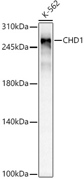
Western blot analysis of lysates from Jurkat cells, using [KO Validated] CHD1 Rabbit pAb (A7883) at 1:500 dilution.
Secondary antibody: HRP Goat Anti-Rabbit IgG (H+L) (AS014) at 1:10000 dilution.
Lysates/proteins: 25μg per lane.
Blocking buffer: 3% nonfat dry milk in TBST.
Detection: ECL Basic Kit (RM00020).
Exposure time: 90s.
Immunohistochemistry analysis of paraffin-embedded Mouse brain using [KO Validated] CHD1 Rabbit pAb (A7883) at dilution of 1:100 (40x lens).Perform microwave antigen retrieval with 10 mM PBS buffer pH 7.2 before commencing with IHC staining protocol.
Immunohistochemistry analysis of paraffin-embedded Human liver damage using [KO Validated] CHD1 Rabbit pAb (A7883) at dilution of 1:100 (40x lens).Perform microwave antigen retrieval with 10 mM PBS buffer pH 7.2 before commencing with IHC staining protocol.
| Product name | [KO Validated] CHD1 Rabbit pAb |
|---|---|
| Catalog No. | A7883 |
| Host species | Rabbit |
| Purification method | Affinity purification |
| Isotype | IgG |
| Immunogen | A synthetic peptide corresponding to a sequence within amino acids 1600 to the C-terminus of human CHD1 (NP_001261.2). |
|---|---|
| Sequence | DREKHRKLDDHRSRDHRSNLEGSLKDRSHSDHRSHSDHRLHSDHRSSSEYTHHKSSRDYRYHSDWQMDHRASSSGPRSPLDQRSPYGSRSPFEHSVEHKSTPEHTWSSRKT |
| Gene ID | |
| Swiss Prot | |
| Synonyms | CHD-1; PILBOS; D1 |
| Calculated MW | 197kDa |
| Observed MW | 250kDa |
| Reactivity | Human, Mouse, Rat |
|---|---|
| Tested applications | WBIHC-PIF/ICCIPChIPChIP-seqRIPFCFC(Intra)ELISAMeDIPNucleotide ArrayDBFACSCoIPCUT&TagmeRIPInhibition |
| Recommended dilution |
|
| Storage buffer | Store at -20℃. Avoid freeze / thaw cycles. Buffer: PBS with 0.02% sodium azide, 50% glycerol, pH7.3. |
| Key application | Western blotting Immunohistochemistry |
| Positive samples | Jurkat |
| Cellular location | Cytoplasm, Nucleus |
| Customer validation | WB(Mus musculus) |
To download a Certificate of Compliance, please enter your Lot number below:
Lot number
* For research use only. Not for therapeutic or diagnostic purposes.
Publishing research using A7883? Please let us know so that we can cite the reference in this datasheet.
![ABclonal:Western blot - [KO Validated] CHD1 Rabbit pAb (A7883)](https://abclonal.oss-cn-hangzhou.aliyuncs.com/A7883_Q1163_KO-WB_01.jpg?t=1715129763 "ABclonal:Western blot - [KO Validated] CHD1 Rabbit pAb (A7883)")
![ABclonal:Western blot - [KO Validated] CHD1 Rabbit pAb (A7883)](https://abclonal.oss-cn-hangzhou.aliyuncs.com/A7883_Q1163_WB_01.jpg?t=1715129763 "ABclonal:Western blot - [KO Validated] CHD1 Rabbit pAb (A7883)")
![ABclonal:Immunohistochemistry - [KO Validated] CHD1 Rabbit pAb (A7883)](https://abclonal.oss-cn-hangzhou.aliyuncs.com/A7883_Q1163_IHC_01.jpg?t=1715129763 "ABclonal:Immunohistochemistry - [KO Validated] CHD1 Rabbit pAb (A7883)")
![ABclonal:Immunohistochemistry - [KO Validated] CHD1 Rabbit pAb (A7883)](https://abclonal.oss-cn-hangzhou.aliyuncs.com/A7883_Q1163_IHC_02.jpg?t=1715129763 "ABclonal:Immunohistochemistry - [KO Validated] CHD1 Rabbit pAb (A7883)")
![ABclonal:Immunohistochemistry - [KO Validated] CHD1 Rabbit pAb (A7883)](https://abclonal.oss-cn-hangzhou.aliyuncs.com/A7883_Q1163_IHC_04.jpg?t=1715129763 "ABclonal:Immunohistochemistry - [KO Validated] CHD1 Rabbit pAb (A7883)")