Tested applications:WBIHC-PIF/ICCIPChIPChIP-seqRIPFCFC(Intra)ELISAMeDIPNucleotide ArrayDBFACSCoIPCUT&TagmeRIPInhibitionReactivity:Human, Mouse, Rat
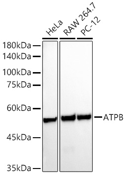
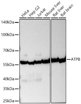
Western blot analysis of various lysates using ATPB Rabbit mAb (A11214) at1:5000 dilution.
Secondary antibody: HRP-conjugated Goat anti-Rabbit IgG (H+L) (AS014) at 1:10000 dilution.
Lysates/proteins: 25μg per lane.
Blocking buffer: 3% nonfat dry milk in TBST.
Detection: ECL Basic Kit (RM00020).
Exposure time: 10s.
Immunohistochemistry analysis of paraffin-embedded Human colon carcinoma tissue using ATPB Rabbit mAb (A11214) at a dilution of 1:200 (40x lens). High pressure antigen retrieval performed with 0.01M Citrate Bufferr (pH 6.0) prior to IHC staining.
Immunohistochemistry analysis of paraffin-embedded Human liver cancer tissue using ATPB Rabbit mAb (A11214) at a dilution of 1:200 (40x lens). High pressure antigen retrieval performed with 0.01M Citrate Bufferr (pH 6.0) prior to IHC staining.
Immunohistochemistry analysis of paraffin-embedded Human liver tissue using ATPB Rabbit mAb (A11214) at a dilution of 1:200 (40x lens). High pressure antigen retrieval performed with 0.01M Citrate Bufferr (pH 6.0) prior to IHC staining.
Immunohistochemistry analysis of paraffin-embedded Human lung adenocarcinoma tissue using ATPB Rabbit mAb (A11214) at a dilution of 1:200 (40x lens). High pressure antigen retrieval performed with 0.01M Citrate Bufferr (pH 6.0) prior to IHC staining.
Immunohistochemistry analysis of paraffin-embedded Human thyroid cancer tissue using ATPB Rabbit mAb (A11214) at a dilution of 1:200 (40x lens). High pressure antigen retrieval performed with 0.01M Citrate Bufferr (pH 6.0) prior to IHC staining.
Immunohistochemistry analysis of paraffin-embedded Mouse kidney tissue using ATPB Rabbit mAb (A11214) at a dilution of 1:200 (40x lens). High pressure antigen retrieval performed with 0.01M Citrate Bufferr (pH 6.0) prior to IHC staining.
Immunohistochemistry analysis of paraffin-embedded Rat kidney tissue using ATPB Rabbit mAb (A11214) at a dilution of 1:200 (40x lens). High pressure antigen retrieval performed with 0.01M Citrate Bufferr (pH 6.0) prior to IHC staining.
Confocal imaging of Hep G2 cells using ATPB Rabbit mAb (A11214, dilution 1:200) followed by a further incubation with Cy3 Goat Anti-Rabbit IgG (H+L) (AS007, dilution 1:500) (Red). The cells were counterstained with α-Tubulin Mouse mAb (AC012, dilution 1:400) followed by incubation with ABflo® 488-conjugated Goat Anti-Mouse IgG (H+L) Ab (AS076, dilution 1:500) (Green). DAPI was used for nuclear staining (Blue). Objective: 100x.
Confocal imaging of NIH/3T3 cells using ATPB Rabbit mAb (A11214, dilution 1:200) followed by a further incubation with Cy3 Goat Anti-Rabbit IgG (H+L) (AS007, dilution 1:500) (Red). The cells were counterstained with α-Tubulin Mouse mAb (AC012, dilution 1:400) followed by incubation with ABflo® 488-conjugated Goat Anti-Mouse IgG (H+L) Ab (AS076, dilution 1:500) (Green). DAPI was used for nuclear staining (Blue). Objective: 100x.
| Product name | ATPB Rabbit mAb |
|---|---|
| Catalog No. | A11214 |
| Host species | Rabbit |
| Purification method | Affinity purification |
| Isotype | IgG |
| CloneNo. | ARC53533 |
| Immunogen | Recombinant fusion protein containing a sequence corresponding to amino acids 230-529 of human ATPB (NP_001677.2). |
|---|---|
| Sequence | YSVFAGVGERTREGNDLYHEMIESGVINLKDATSKVALVYGQMNEPPGARARVALTGLTVAEYFRDQEGQDVLLFIDNIFRFTQAGSEVSALLGRIPSAVGYQPTLATDMGTMQERITTTKKGSITSVQAIYVPADDLTDPAPATTFAHLDATTVLSRAIAELGIYPAVDPLDSTSRIMDPNIVGSEHYDVARGVQKILQDYKSLQDIIAILGMDELSEEDKLTVSRARKIQRFLSQPFQVAEVFTGHMGKLVPLKETIKGFQQILAGEYDHLPEQAFYMVGPIEEAVAKADKLAEEHSS |
| Gene ID | |
| Swiss Prot | |
| Synonyms | ATP5B; ATPMB; ATPSB; HUMOP2; HEL-S-271; ATPB |
| Calculated MW | 57kDa |
| Observed MW | 57kDa |
| Reactivity | Human, Mouse, Rat |
|---|---|
| Tested applications | WBIHC-PIF/ICCIPChIPChIP-seqRIPFCFC(Intra)ELISAMeDIPNucleotide ArrayDBFACSCoIPCUT&TagmeRIPInhibition |
| Recommended dilution |
|
| Storage buffer | Store at -20℃. Avoid freeze / thaw cycles. Buffer: PBS with 0.05% proclin300, 0.05% BSA, 50% glycerol, pH7.3. |
| Key application | Western blotting Immunohistochemistry Immunofluorescence |
| Positive samples | HeLa, RAW 264.7, PC-12, Mouse heart |
| Cellular location | Mitochondrion, Mitochondrion inner membrane. |
To download a Certificate of Compliance, please enter your Lot number below:
Lot number
* For research use only. Not for therapeutic or diagnostic purposes.
")
")
")
")
")
")
")
")
")
")