Tested applications:WBIHC-PIF/ICCIPChIPChIP-seqRIPFCFC(Intra)ELISAMeDIPNucleotide ArrayDBFACSCoIPCUT&TagmeRIPInhibitionReactivity:Human
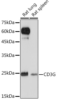
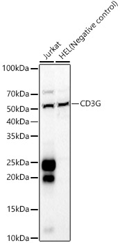
Western blot analysis of various lysates using CD3G Rabbit mAb (A25012) at 1:3000 dilution.
Secondary antibody: HRP Goat Anti-Rabbit IgG (H+L) (AS014) at 1:10000 dilution.
Lysates/proteins: 25ug per lane.
Blocking buffer: 3% nonfat dry milk in TBST.
Detection: ECL Basic Kit (RM00020).
Negative control (NC):HEL
Exposure time: 20s.
Immunohistochemistry analysis of CD3G in paraffin-embedded human colon tissue using CD3G Rabbit mAb (A25012) at a dilution of 1:800 (40x lens).High pressure antigen retrieval was performed with 0.01 M citrate buffer (pH 6.0) prior to IHC staining.
Immunohistochemistry analysis of CD3G in paraffin-embedded human tonsil tissue using CD3G Rabbit mAb (A25012) at a dilution of 1:800 (40x lens).High pressure antigen retrieval was performed with 0.01 M citrate buffer (pH 6.0) prior to IHC staining.
Confocal imaging of Jurkat cells using CD3G Rabbit mAb (A25012, dilution 1:200) followed by a further incubation with Cy3 Goat Anti-Rabbit IgG (H+L) (AS007, dilution 1:500) (Red). DAPI was used for nuclear staining (Blue). Objective: 100x.
| Product name | CD3G Rabbit mAb |
|---|---|
| Catalog No. | A25012 |
| Host species | Rabbit |
| Purification method | Affinity purification |
| Isotype | IgG |
| CloneNo. | ARC65124 |
| Immunogen | Recombinant fusion protein containing a sequence corresponding to amino acids 23-116 of human CD3G (NP_000064.1). |
|---|---|
| Sequence | QSIKGNHLVKVYDYQEDGSVLLTCDAEAKNITWFKDGKMIGFLTEDKKKWNLGSNAKDPRGMYQCKGSQNKSKPLQVYYRMCQNCIELNAATIS |
| Gene ID | |
| Swiss Prot | |
| Synonyms | T3G; IMD17; CD3GAMMA; CD3-GAMMA; CD3G |
| Calculated MW | 20kDa |
| Observed MW | 18-28kDa |
| Reactivity | Human |
|---|---|
| Tested applications | WBIHC-PIF/ICCIPChIPChIP-seqRIPFCFC(Intra)ELISAMeDIPNucleotide ArrayDBFACSCoIPCUT&TagmeRIPInhibition |
| Recommended dilution |
|
| Storage buffer | Store at -20℃. Avoid freeze / thaw cycles. Buffer: PBS with 0.05% proclin300, 0.05% BSA, 50% glycerol, pH7.3. |
| Key application | Western blotting Immunohistochemistry Immunofluorescence |
| Positive samples | MOLT-4, Jurkat |
| Cellular location | Cell membrane , Single-pass type I membrane protein |
| Customer validation | WB(Homo sapiens, Mus musculus) Co-IP(Homo sapiens, Mus musculus) RT-PCR(Homo sapiens, Mus musculus) ChIP(Homo sapiens) Co-IP(Homo sapiens) FC(Homo sapiens) IF(Homo sapiens) RT-qPCR(Homo sapiens) |
To download a Certificate of Compliance, please enter your Lot number below:
Lot number
* For research use only. Not for therapeutic or diagnostic purposes.
Publishing research using A25012? Please let us know so that we can cite the reference in this datasheet.
")
")
")
")
")