Tested applications:WBIHC-PIF/ICCIPChIPChIP-seqRIPFCFC(Intra)ELISAMeDIPNucleotide ArrayDBFACSCoIPCUT&TagmeRIPInhibitionReactivity:Human, Mouse, Rat
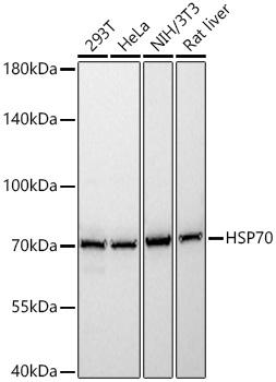
Western blot analysis of various lysates using HSP70 Rabbit pAb (A20819) at 1:1000 dilution.
Secondary antibody: HRP-conjugated Goat anti-Rabbit IgG (H+L) (AS014) at 1:10000 dilution.
Lysates/proteins: 25μg per lane.
Blocking buffer: 3% nonfat dry milk in TBST.
Detection: ECL Basic Kit (RM00020).
Exposure time: 0.5s.
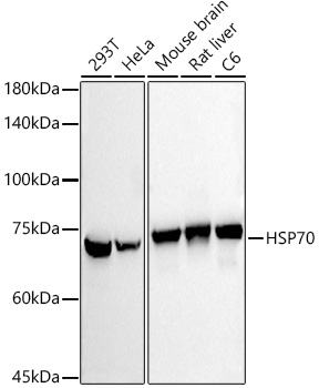
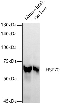
Western blot analysis of various lysates using HSP70 Rabbit pAb (A20819) at 1:1000 dilution.
Secondary antibody: HRP-conjugated Goat anti-Rabbit IgG (H+L) (AS014) at 1:10000 dilution.
Lysates/proteins: 25μg per lane.
Blocking buffer: 3% nonfat dry milk in TBST.
Detection: ECL Basic Kit (RM00020).
Exposure time: 10s.
Immunohistochemistry analysis of paraffin-embedded Human colon carcinoma using HSP70 Rabbit pAb (A20819) at dilution of 1:100 (40x lens). High pressure antigen retrieval performed with 0.01M Citrate Bufferr (pH 6.0) prior to IHC staining.
Immunohistochemistry analysis of paraffin-embedded Human esophageal cancer using HSP70 Rabbit pAb (A20819) at dilution of 1:100 (40x lens). High pressure antigen retrieval performed with 0.01M Citrate Bufferr (pH 6.0) prior to IHC staining.
Immunofluorescence analysis of A-549 cells using HSP70 Rabbit pAb (A20819) at dilution of 1:100 (40x lens). Secondary antibody: Cy3-conjugated Goat anti-Rabbit IgG (H+L) (AS007) at 1:500 dilution. Blue: DAPI for nuclear staining.
Immunofluorescence analysis of HeLa cells using HSP70 Rabbit pAb (A20819) at dilution of 1:100 (40x lens). Secondary antibody: Cy3-conjugated Goat anti-Rabbit IgG (H+L) (AS007) at 1:500 dilution. Blue: DAPI for nuclear staining.
Immunofluorescence analysis of NIH/3T3 cells using HSP70 Rabbit pAb (A20819) at dilution of 1:100 (40x lens). Secondary antibody: Cy3-conjugated Goat anti-Rabbit IgG (H+L) (AS007) at 1:500 dilution. Blue: DAPI for nuclear staining.
Immunofluorescence analysis of PC-12 cells using HSP70 Rabbit pAb (A20819) at dilution of 1:100 (40x lens). Secondary antibody: Cy3-conjugated Goat anti-Rabbit IgG (H+L) (AS007) at 1:500 dilution. Blue: DAPI for nuclear staining.
| Product name | HSP70 Rabbit pAb |
|---|---|
| Catalog No. | A20819 |
| Host species | Rabbit |
| Purification method | Affinity purification |
| Isotype | IgG |
| Immunogen | A synthetic peptide corresponding to a sequence within amino acids 500-600 of human HSP70 (NP_005336.3). |
|---|---|
| Sequence | KITITNDKGRLSKEEIERMVQEAEKYKAEDEVQRERVSAKNALESYAFNMKSAVEDEGLKGKISEADKKKVLDKCQEVISWLDANTLAEKDEFEHKRKELE |
| Gene ID | |
| Swiss Prot | |
| Synonyms | HEL-S-103; HSP70-1; HSP70-1A; HSP70.1; HSP70I; HSP72; HSPA1; HSP70 |
| Calculated MW | 70kDa |
| Observed MW | 70kDa |
| Reactivity | Human, Mouse, Rat |
|---|---|
| Tested applications | WBIHC-PIF/ICCIPChIPChIP-seqRIPFCFC(Intra)ELISAMeDIPNucleotide ArrayDBFACSCoIPCUT&TagmeRIPInhibition |
| Recommended dilution |
|
| Storage buffer | Store at -20℃. Avoid freeze / thaw cycles. Buffer: PBS with 0.05% proclin300, 50% glycerol, pH7.3. |
| Key application | Western blotting Immunohistochemistry Immunofluorescence |
| Positive samples | 293T, HeLa, A-549, NIH/3T3, C6, Mouse brain, Mouse lung, Rat heart, Rat liver |
| Cellular location | blood microparticle, centriole, centrosome, cytoplasm, cytosol, endoplasmic reticulum, extracellular exosome, extracellular region, focal adhesion, mitochondrion, nuclear speck, nucleoplasm, nucleus, perinuclear region of cytoplasm, plasma membrane. |
| Customer validation | WB(Gallus gallus, Other) IF(Mus musculus) |
To download a Certificate of Compliance, please enter your Lot number below:
Lot number
* For research use only. Not for therapeutic or diagnostic purposes.
Publishing research using A20819? Please let us know so that we can cite the reference in this datasheet.
")
")
")
")
")
")
")
")
")