Review (1)Publication (1) Datasheet
Tested applications:WBIHC-PIF/ICCIPChIPChIP-seqRIPFCFC(Intra)ELISAMeDIPNucleotide ArrayDBFACSCoIPCUT&TagmeRIPInhibitionReactivity:Human, Mouse, Rat
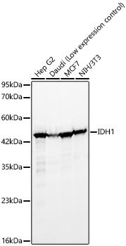
Western blot analysis of various lysates, using IDH1 Rabbit mAb (A5106) at 1:10000 dilution.
Secondary antibody: HRP-conjugated Goat anti-Rabbit IgG (H+L) (AS014) at 1:10000 dilution.
Lysates/proteins: 25μg per lane.
Blocking buffer: 3% nonfat dry milk in TBST.
Detection: ECL Basic Kit (RM00020).
Exposure time: 90s.
Confocal imaging of NIH/3T3 cells using IDH1 Rabbit mAb (A5106, dilution1:100) (Red). The cells were counterstained with α-Tubulin (AC012, dilution 1:400)(Green). DAPI was used for nuclear staining (blue). Objective: 60x
| Product name | IDH1 Rabbit mAb |
|---|---|
| Catalog No. | A5106 |
| Host species | Rabbit |
| Purification method | Affinity purification |
| Isotype | IgG |
| CloneNo. | ARC54138 |
| Immunogen | A synthetic peptide corresponding to a sequence within amino acids 100-200 of human IDH1 (NP_005887.2). |
|---|---|
| Sequence | RNILGGTVFREAIICKNIPRLVSGWVKPIIIGRHAYGDQYRATDFVVPGPGKVEITYTPSDGTQKVTYLVHNFEEGGGVAMGMYNQDKSIEDFAHSSFQMA |
| Gene ID | |
| Swiss Prot | |
| Synonyms | IDH; IDP; IDCD; IDPC; PICD; HEL-216; HEL-S-26; IDH1 |
| Calculated MW | 47kDa |
| Observed MW | 46kDa |
| Reactivity | Human, Mouse, Rat |
|---|---|
| Tested applications | WBIHC-PIF/ICCIPChIPChIP-seqRIPFCFC(Intra)ELISAMeDIPNucleotide ArrayDBFACSCoIPCUT&TagmeRIPInhibition |
| Recommended dilution |
|
| Storage buffer | Store at -20℃. Avoid freeze / thaw cycles. Buffer: PBS with 0.05% proclin300, 0.05% BSA, 50% glycerol, pH7.3. |
| Key application | Western blotting Immunofluorescence |
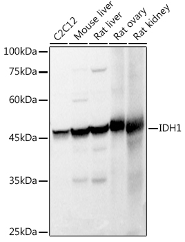
| Positive samples | Hep G2, MCF7, 293F, NIH/3T3, C2C12 |
| Cellular location | Cytoplasm, Peroxisome. |
| Customer validation | (HeLa、NCL-H460、U-87mg、MCF-7、HepG2) WB(Mus musculus) |
To download a Certificate of Compliance, please enter your Lot number below:
Lot number
* For research use only. Not for therapeutic or diagnostic purposes.
Please submit reviews to your technical sales specialist directly. Alternatively, email us at info@abclonal.co.kr
Publishing research using A5106? Please let us know so that we can cite the reference in this datasheet.
")
")
")
![[KO Validated] IDH1 Rabbit pAb](https://abclonal.oss-cn-hangzhou.aliyuncs.com/A13245_N216-4_KO-WB_01.jpg?t=1709113168)