Tested applications:WBIHC-PIF/ICCIPChIPChIP-seqRIPFCFC(Intra)ELISAMeDIPNucleotide ArrayDBFACSCoIPCUT&TagmeRIPInhibitionReactivity:Human, Mouse, Rat
Immunohistochemistry analysis of paraffin-embedded Human colon tissue using Ki67 Rabbit mAb (A23722) at a dilution of 1:1300 (40x lens). High pressure antigen retrieval performed with 0.01M Citrate Buffer (pH 6.0) prior to IHC staining.
Immunohistochemistry analysis of paraffin-embedded Human tonsil tissue using Ki67 Rabbit mAb (A23722) at a dilution of 1:1300 (40x lens). High pressure antigen retrieval performed with 0.01M Citrate Buffer (pH 6.0) prior to IHC staining.
Immunohistochemistry analysis of paraffin-embedded Mouse colon tissue using Ki67 Rabbit mAb (A23722) at a dilution of 1:1300 (40x lens). High pressure antigen retrieval performed with 0.01M Citrate Buffer (pH 6.0) prior to IHC staining.
Immunohistochemistry analysis of paraffin-embedded Mouse spleen tissue using Ki67 Rabbit mAb (A23722) at a dilution of 1:1300 (40x lens). High pressure antigen retrieval performed with 0.01M Citrate Buffer (pH 6.0) prior to IHC staining.
Immunohistochemistry analysis of paraffin-embedded Mouse testis tissue using Ki67 Rabbit mAb (A23722) at a dilution of 1:1300 (40x lens). High pressure antigen retrieval performed with 0.01M Citrate Buffer (pH 6.0) prior to IHC staining.
Immunohistochemistry analysis of paraffin-embedded Rat colon tissue using Ki67 Rabbit mAb (A23722) at a dilution of 1:1300 (40x lens). High pressure antigen retrieval performed with 0.01M Citrate Buffer (pH 6.0) prior to IHC staining.
Immunohistochemistry analysis of paraffin-embedded Rat spleen tissue using Ki67 Rabbit mAb (A23722) at a dilution of 1:1300 (40x lens). High pressure antigen retrieval performed with 0.01M Citrate Buffer (pH 6.0) prior to IHC staining.
Confocal imaging of HeLa cells using Ki67 Rabbit mAb (A23722, dilution 1:200) followed by a further incubation with Cy3 Goat Anti-Rabbit IgG (H+L) (AS007, dilution 1:500) (Red). The cells were counterstained with α-Tubulin Mouse mAb (AC012, dilution 1:400) followed by incubation with ABflo® 488-conjugated Goat Anti-Mouse IgG (H+L) Ab (AS076, dilution 1:500) (Green). DAPI was used for nuclear staining (Blue). Objective: 100x.
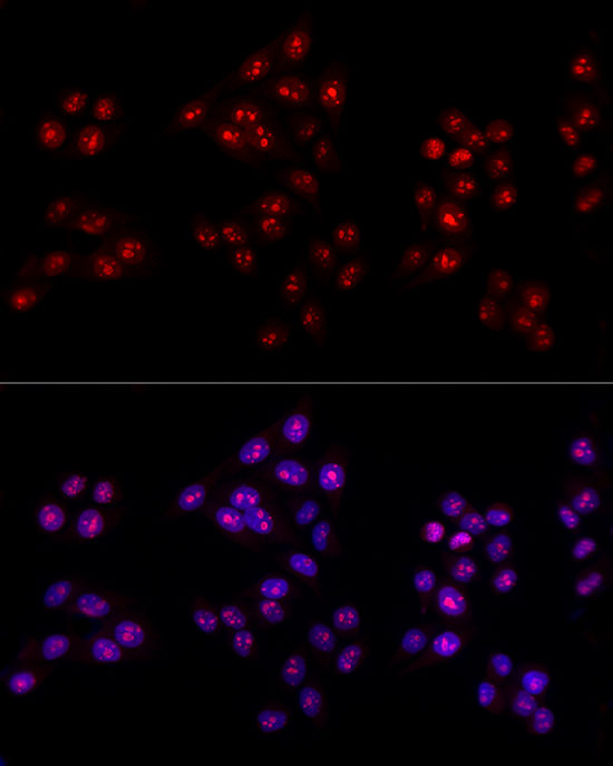
Confocal imaging of Neuro-2a cells using Ki67 Rabbit mAb (A23722, dilution 1:200) followed by a further incubation with Cy3 Goat Anti-Rabbit IgG (H+L) (AS007, dilution 1:500) (Red). The cells were counterstained with α-Tubulin Mouse mAb (AC012, dilution 1:400) followed by incubation with ABflo® 488-conjugated Goat Anti-Mouse IgG (H+L) Ab (AS076, dilution 1:500) (Green). DAPI was used for nuclear staining (Blue). Objective: 100x.
Confocal imaging of paraffin-embedded Rat spleen tissue using Ki67 Rabbit mAb (A23722, dilution 1:200) followed by a further incubation with Cy3 Goat Anti-Rabbit IgG (H+L) (AS007, dilution 1:500) (Red). DAPI was used for nuclear staining (Blue). High pressure antigen retrieval performed with 0.01M Citrate Buffer (pH 6.0) prior to IF staining. Objective: 40x.
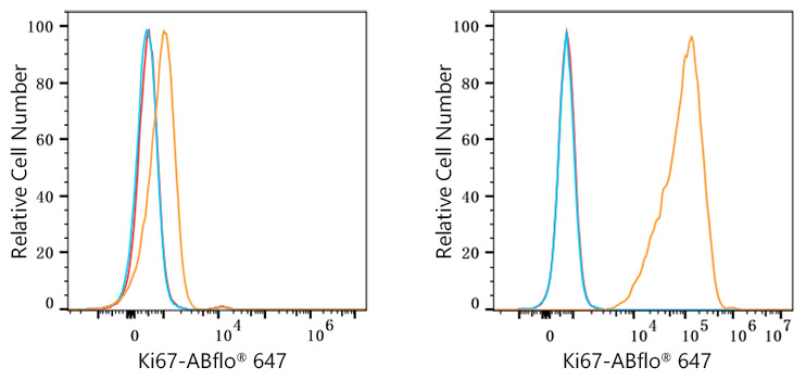
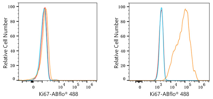
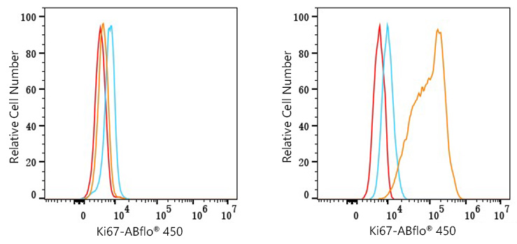
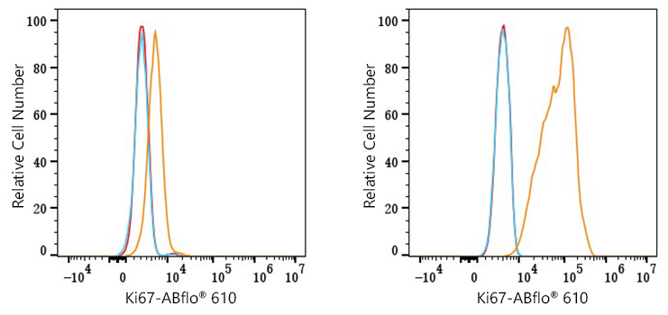
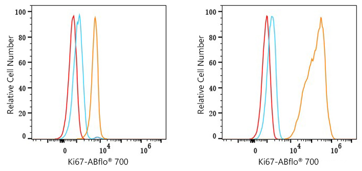
Flow cytometry: 1X10^6 knockout (KO) HeLa cells (negative control, left) and HeLa cells (right) were intracellularly-stained with Ki67 Rabbit mAb (A23722, 2 μg/mL, orange line) or Rabbit IgG isotype control (AC042, 2 μg/mL, blue line), followed by FITC conjugated goat anti-Rabbit pAb staining. Non-fluorescently stained cells were used as blank control (red line).
| Product name | Ki67 Rabbit mAb |
|---|---|
| Catalog No. | A23722 |
| Host species | Rabbit |
| Purification method | Affinity purification |
| Isotype | IgG |
| CloneNo. | ARC57561 |
| Immunogen | A synthetic peptide corresponding to a sequence within amino acids 2271-2370 of human Ki67 (NP_002408.3). |
|---|---|
| Sequence | PAGGDEKDMKAFMGTPVQKLDLPGNLPGSKRWPQTPKEKAQALEDLAGFKELFQTPGTDKPTTDEKTTKIACKSPQPDPVDTPASTKQRPKRNLRKADVE |
| Gene ID | |
| Swiss Prot | |
| Synonyms | KIA; MIB-; MIB-1; PPP1R105; Ki67 |
| Calculated MW | 359kDa |
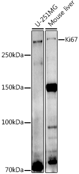
| Observed MW | Refer to figures |
| Reactivity | Human, Mouse, Rat |
|---|---|
| Tested applications | WBIHC-PIF/ICCIPChIPChIP-seqRIPFCFC(Intra)ELISAMeDIPNucleotide ArrayDBFACSCoIPCUT&TagmeRIPInhibition |
| Recommended dilution |
|
| Storage buffer | Store at -20℃. Avoid freeze / thaw cycles. Buffer: PBS with 0.05% proclin300, 0.05% BSA, 50% glycerol, pH7.3. |
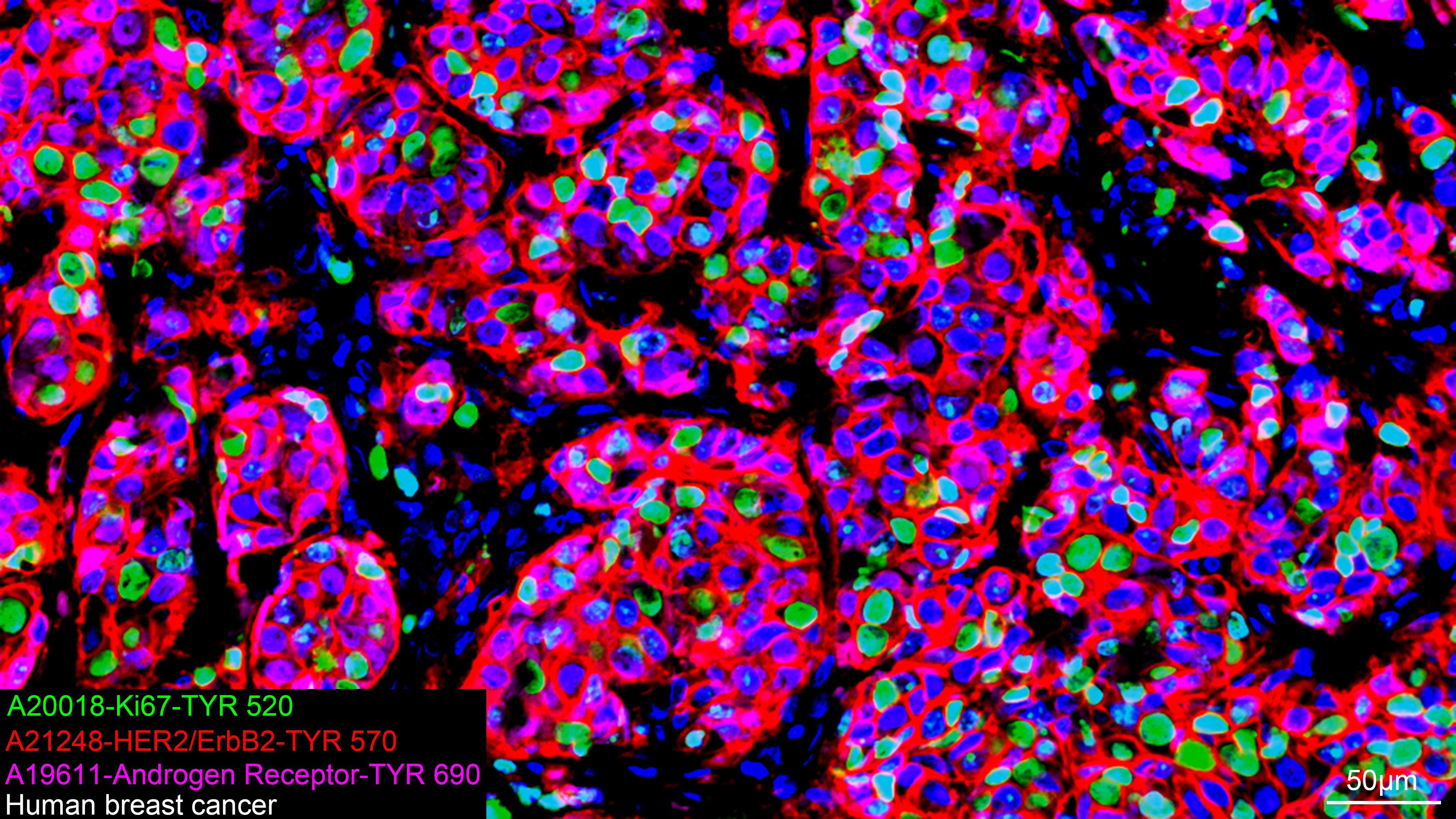
| Key application | Immunohistochemistry Immunofluorescence Flow Cytometry |
| Positive samples | |
| Cellular location | Chromosome, Nucleus, nucleolus. |
| Customer validation | IF(Rattus norvegicus) IHC(Mus musculus, Homo sapiens) Other(Homo sapiens) WB(Homo sapiens) |
To download a Certificate of Compliance, please enter your Lot number below:
Lot number
* For research use only. Not for therapeutic or diagnostic purposes.
Publishing research using A23722? Please let us know so that we can cite the reference in this datasheet.
")
")
")
")
")
")
")
")
")
")
")
")