Tested applications:WBIHC-PIF/ICCIPChIPChIP-seqRIPFCFC(Intra)ELISAMeDIPNucleotide ArrayDBFACSCoIPCUT&TagmeRIPInhibitionReactivity:Human, Mouse
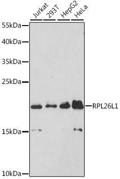
Western blot analysis of various lysates using (A9419) at 1:1000 dilution.
Secondary antibody: HRP Goat Anti-Rabbit IgG (H+L) (AS014) at 1:10000 dilution.
Lysates/proteins: 25μg per lane.
Blocking buffer: 3% nonfat dry milk in TBST.
Detection: ECL Basic Kit (RM00020).
Exposure time: 3s.
Immunohistochemistry analysis of paraffin-embedded Human lung cancer using RPL26L1 Rabbit mAb (A9419) at dilution of 1:50 (40x lens). Perform high pressure antigen retrieval with 10 mM citrate buffer pH 6.0 before commencing with IHC staining protocol.
Immunohistochemistry analysis of paraffin-embedded Human tonsil using RPL26L1 Rabbit mAb (A9419) at dilution of 1:50 (40x lens). Perform high pressure antigen retrieval with 10 mM citrate buffer pH 6.0 before commencing with IHC staining protocol.
Confocal imaging of paraffin-embedded Mouse brain tissue using RPL26L1 Rabbit mAb (A9419, dilution 1:100) followed by a further incubation with Cy3 Goat Anti-Rabbit IgG (H+L) (AS007, dilution 1:500) (Red). DAPI was used for nuclear staining (Blue). Microwave antigen retrieval performed with 0.01M Citrate Buffer(pH 6.0) prior to IF staining. Objective: 40x.
Confocal imaging of U-2 OS cells using RPL26L1 Rabbit mAb (A9419, dilution 1:50) followed by a further incubation with Cy3 Goat Anti-Rabbit IgG (H+L) (AS007, dilution 1:500) (Red). The cells were counterstained with α-Tubulin Mouse mAb (AC012, dilution 1:400) followed by incubation with ABflo® 488-conjugated Goat Anti-Mouse IgG (H+L) Ab (AS076, dilution 1:500) (Green). DAPI was used for nuclear staining (Blue). Objective: 100x.
Confocal imaging of MCF7 cells using RPL26L1 Rabbit mAb (A9419, dilution 1:50) followed by a further incubation with Cy3 Goat Anti-Rabbit IgG (H+L) (AS007, dilution 1:500) (Red). The cells were counterstained with α-Tubulin Mouse mAb (AC012, dilution 1:400) followed by incubation with ABflo® 488-conjugated Goat Anti-Mouse IgG (H+L) Ab (AS076, dilution 1:500) (Green). DAPI was used for nuclear staining (Blue). Objective: 100x.
| Product name | RPL26L1 Rabbit mAb |
|---|---|
| Catalog No. | A9419 |
| Host species | Rabbit |
| Purification method | Affinity purification |
| Isotype | IgG |
| CloneNo. | ARC2783 |
| Immunogen | A synthetic peptide corresponding to a sequence within amino acids 1-100 of human RPL26L1 (Q9UNX3). |
|---|---|
| Sequence | MKFNPFVTSDRSKNRKRHFNAPSHVRRKIMSSPLSKELRQKYNVRSMPIRKDDEVQVVRGHYKGQQIGKVVQVYRKKYVIYIERVQREKANGTTVHVGIH |
| Gene ID | |
| Swiss Prot | |
| Synonyms | RPL26P1; RPL26L1 |
| Calculated MW | 17kDa |
| Observed MW | 20kDa |
| Reactivity | Human, Mouse |
|---|---|
| Tested applications | WBIHC-PIF/ICCIPChIPChIP-seqRIPFCFC(Intra)ELISAMeDIPNucleotide ArrayDBFACSCoIPCUT&TagmeRIPInhibition |
| Recommended dilution |
|
| Storage buffer | Store at -20℃. Avoid freeze / thaw cycles. Buffer: PBS with 0.02% sodium azide, 0.05% BSA, 50% glycerol, pH7.3. |
| Key application | Western blotting Immunohistochemistry Immunofluorescence |
| Positive samples | HeLa, 293T, HepG2, A-549 |
| Cellular location | extracellular exosome |
To download a Certificate of Compliance, please enter your Lot number below:
Lot number
* For research use only. Not for therapeutic or diagnostic purposes.
")
")
")
")
")
")