Tested applications:WBIHC-PIF/ICCIPChIPChIP-seqRIPFCFC(Intra)ELISAMeDIPNucleotide ArrayDBFACSCoIPCUT&TagmeRIPInhibitionReactivity:Mouse
The multiplex IHC analysis on paraffin-embedded Mouse brain tissue using the following specific primary antibodies and tyramide signal amplification (TSA) reagents (RK05903) : NeuN Rabbit mAb (A19086, 1:2000) with TSA-TYR-520 (Green), GFAP Rabbit mAb (A19058, 1:500) with TSA-TYR-570 (Red), and TMEM119 Rabbit mAb (A27143, 1:600) with TSA-TYR-690 (Yellow). DAPI (Blue) was used for nuclear staining. Prior to multiplex IHC staining, high-pressure antigen retrieval was performed using 0.01M citrate buffer at pH 6.0. The analysis was completed using a 20x objective lens.
The multiplex IHC analysis on paraffin-embedded Rat brain tissue using the following specific primary antibodies and tyramide signal amplification (TSA) reagents (RK05903) : NeuN Rabbit mAb (A19086, 1:2000) with TSA-TYR-520 (Green), GFAP Rabbit mAb (A19058, 1:500) with TSA-TYR-570 (Red), and TMEM119 Rabbit mAb (A27143, 1:600) with TSA-TYR-690 (Yellow). DAPI (Blue) was used for nuclear staining. Prior to multiplex IHC staining, high-pressure antigen retrieval was performed using 0.01M citrate buffer at pH 6.0. The analysis was completed using a 20x objective lens.
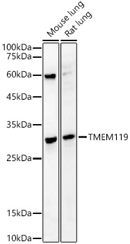
Western blot analysis of lysates from Mouse brain using TMEM119 Rabbit mAb (A27143) at 1:12000 dilution incubated overnight at 4℃.
Secondary antibody: HRP-conjugated Goat anti-Rabbit IgG (H+L) (AS014) at 1:10000 dilution.
Lysates/proteins: 25 μg per lane.
Blocking buffer: 3% nonfat dry milk in TBST.
Detection: ECL Basic Kit (RM00020).
Exposure time: 45s.
Immunohistochemistry analysis of paraffin-embedded Mouse brain tissue using TMEM119 Rabbit mAb (A27143) at a dilution of 1:6000 (40x lens). High pressure antigen retrieval performed with 0.01M Tris-EDTA Buffer (pH 9.0) prior to IHC staining.
Immunohistochemistry analysis of paraffin-embedded Mouse intestine tissue using TMEM119 Rabbit mAb (A27143) at a dilution of 1:6000 (40x lens). High pressure antigen retrieval performed with 0.01M Tris-EDTA Buffer (pH 9.0) prior to IHC staining.
Confocal imaging of paraffin-embedded Mouse brain tissue using TMEM119 Rabbit mAb (A27143, dilution 1:500) followed by a further incubation with Cy3 Goat Anti-Rabbit IgG (H+L) (AS007, dilution 1:500) (Red). DAPI was used for nuclear staining (Blue). High pressure antigen retrieval performed with 0.01M Citrate Buffer (pH 6.0) prior to IF staining. Objective: 40x.
| Product name | TMEM119 Rabbit mAb |
|---|---|
| Catalog No. | A27143 |
| Host species | Rabbit |
| Purification method | Affinity purification |
| Isotype | IgG |
| CloneNo. | ARC5161 |
| Immunogen | Recombinant protein of human TMEM119. |
|---|---|
| Sequence | Email for sequence |
| Gene ID | |
| Swiss Prot | |
| Synonyms | OBIF |
| Calculated MW | 29kDa |
| Observed MW | 56kDa |
| Reactivity | Mouse |
|---|---|
| Tested applications | WBIHC-PIF/ICCIPChIPChIP-seqRIPFCFC(Intra)ELISAMeDIPNucleotide ArrayDBFACSCoIPCUT&TagmeRIPInhibition |
| Recommended dilution |
|
| Storage buffer | Store at -20℃. Avoid freeze / thaw cycles. Buffer: PBS with 0.09% Sodium azide, 0.05% BSA, 50% glycerol, pH7.3. |
| Key application | Western blotting Immunohistochemistry Immunofluorescence |
| Positive samples | Mouse brain |
| Cellular location | plasma membrane. |
| Customer validation | IF(Mus musculus) WB(Mus musculus) |
To download a Certificate of Compliance, please enter your Lot number below:
Lot number
* For research use only. Not for therapeutic or diagnostic purposes.
Publishing research using A27143? Please let us know so that we can cite the reference in this datasheet.
")
")
")
")
")
")