Tested applications:WBIHC-PIF/ICCIPChIPChIP-seqRIPFCFC(Intra)ELISAMeDIPNucleotide ArrayDBFACSCoIPCUT&TagmeRIPInhibitionReactivity:Human, Mouse, Rat
Immunohistochemistry analysis of paraffin-embedded Human esophagus tissue using mSin3A Rabbit mAb (A2396) at a dilution of 1:200 (40x lens). High pressure antigen retrieval was performed with 0.01 M citrate buffer (pH 6.0) prior to IHC staining.
Immunohistochemistry analysis of paraffin-embedded Human small intestine tissue using mSin3A Rabbit mAb (A2396) at a dilution of 1:200 (40x lens). High pressure antigen retrieval was performed with 0.01 M citrate buffer (pH 6.0) prior to IHC staining.
Immunohistochemistry analysis of paraffin-embedded Mouse brain tissue using mSin3A Rabbit mAb (A2396) at a dilution of 1:200 (40x lens). High pressure antigen retrieval was performed with 0.01 M citrate buffer (pH 6.0) prior to IHC staining.
Immunohistochemistry analysis of paraffin-embedded Mouse colon tissue using mSin3A Rabbit mAb (A2396) at a dilution of 1:200 (40x lens). High pressure antigen retrieval was performed with 0.01 M citrate buffer (pH 6.0) prior to IHC staining.
Immunohistochemistry analysis of paraffin-embedded Mouse testis tissue using mSin3A Rabbit mAb (A2396) at a dilution of 1:200 (40x lens). High pressure antigen retrieval was performed with 0.01 M citrate buffer (pH 6.0) prior to IHC staining.
Immunohistochemistry analysis of paraffin-embedded Rat colon tissue using mSin3A Rabbit mAb (A2396) at a dilution of 1:200 (40x lens). High pressure antigen retrieval was performed with 0.01 M citrate buffer (pH 6.0) prior to IHC staining.
Immunohistochemistry analysis of paraffin-embedded Rat spleen tissue using mSin3A Rabbit mAb (A2396) at a dilution of 1:200 (40x lens). High pressure antigen retrieval was performed with 0.01 M citrate buffer (pH 6.0) prior to IHC staining.
Immunohistochemistry analysis of paraffin-embedded Rat testis tissue using mSin3A Rabbit mAb (A2396) at a dilution of 1:200 (40x lens). High pressure antigen retrieval was performed with 0.01 M citrate buffer (pH 6.0) prior to IHC staining.
Confocal imaging of NIH/3T3 cells using mSin3A Rabbit mAb (A2396, dilution 1:200) followed by a further incubation with Cy3 Goat Anti-Rabbit IgG (H+L) (AS007, dilution 1:500) (Red). The cells were counterstained with α-Tubulin Mouse mAb (AC012, dilution 1:400) followed by incubation with ABflo® 488-conjugated Goat Anti-Mouse IgG (H+L) Ab (AS076, dilution 1:500) (Green). DAPI was used for nuclear staining (Blue). Objective: 100x.
Confocal imaging of U-2 OS cells using mSin3A Rabbit mAb (A2396, dilution 1:200) followed by a further incubation with Cy3 Goat Anti-Rabbit IgG (H+L) (AS007, dilution 1:500) (Red). The cells were counterstained with α-Tubulin Mouse mAb (AC012, dilution 1:400) followed by incubation with ABflo® 488-conjugated Goat Anti-Mouse IgG (H+L) Ab (AS076, dilution 1:500) (Green). DAPI was used for nuclear staining (Blue). Objective: 100x.
| Product name | mSin3A Rabbit mAb |
|---|---|
| Catalog No. | A2396 |
| Host species | Rabbit |
| Purification method | Affinity purification |
| Isotype | IgG |
| CloneNo. | ARC0745 |
| Immunogen | A synthetic peptide corresponding to a sequence within amino acids 1174-1273 of human mSin3A (Q96ST3). |
|---|---|
| Sequence | YKMVYVIKSEDYMYRRTALLRAHQSHERVSKRLHQRFQAWVDKWTKEHVPREMAAETSKWLMGEGLEGLVPCTTTCDTETLHFVSINKYRVKYGTVFKAP |
| Gene ID | |
| Swiss Prot | |
| Synonyms | WITKOS; DEL15Q24; CHR15DELq24; mSin3A |
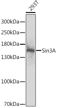
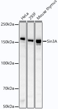
| Calculated MW | 145kDa |
| Observed MW | Refer to figures |
| Reactivity | Human, Mouse, Rat |
|---|---|
| Tested applications | WBIHC-PIF/ICCIPChIPChIP-seqRIPFCFC(Intra)ELISAMeDIPNucleotide ArrayDBFACSCoIPCUT&TagmeRIPInhibition |
| Recommended dilution |
|
| Storage buffer | Store at -20℃. Avoid freeze / thaw cycles. Buffer: PBS with 0.02% sodium azide, 0.05% BSA, 50% glycerol, pH7.3. |
| Key application | Immunohistochemistry Immunofluorescence |
| Positive samples | |
| Cellular location | histone deacetylase complex, nucleolus, nucleoplasm, nucleus. |
To download a Certificate of Compliance, please enter your Lot number below:
Lot number
* For research use only. Not for therapeutic or diagnostic purposes.
")
")
")
")
")
")
")
")
")
")