Tested applications:WBIHC-PIF/ICCIPChIPChIP-seqRIPFCFC(Intra)ELISAMeDIPNucleotide ArrayDBFACSCoIPCUT&TagmeRIPInhibitionReactivity:Human, Mouse, Rat
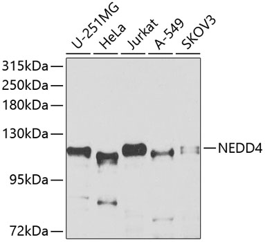
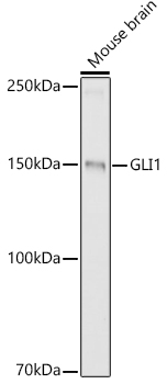
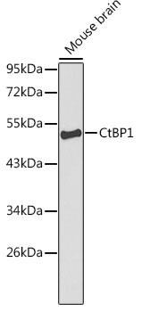
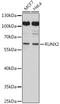
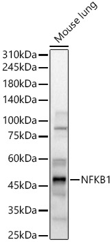
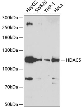
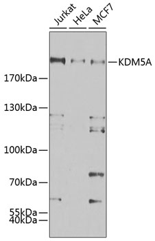
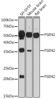
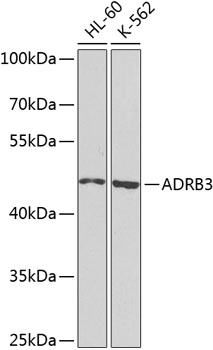
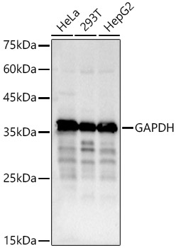
Western blot analysis of various lysates using ASH2L Rabbit mAb (A4892) at 1:1000 dilution.
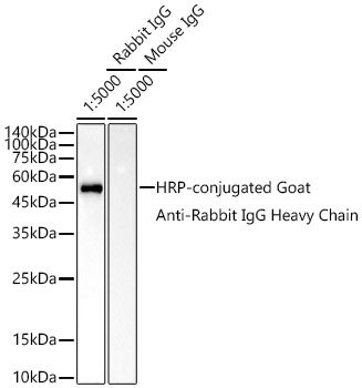
Secondary antibody: HRP Goat Anti-Rabbit IgG (H+L) (AS014) at 1:10000 dilution.
Lysates/proteins: 25μg per lane.
Blocking buffer: 3% nonfat dry milk in TBST.
Detection: ECL Basic Kit (RM00020).
Exposure time: 5s.
Immunohistochemistry analysis of ASH2L in paraffin-embedded mouse lung tissue using ASH2L Rabbit mAb (A4892) at a dilution of 1:200 (40x lens). High pressure antigen retrieval was performed with 0.01 M citrate buffer (pH 6.0) prior to IHC staining.
Immunohistochemistry analysis of ASH2L in paraffin-embedded rat brain tissue using ASH2L Rabbit mAb (A4892) at a dilution of 1:200 (40x lens). High pressure antigen retrieval was performed with 0.01 M citrate buffer (pH 6.0) prior to IHC staining.
Immunohistochemistry analysis of ASH2L in paraffin-embedded human colon carcinoma tissue using ASH2L Rabbit mAb (A4892) at a dilution of 1:200 (40x lens). High pressure antigen retrieval was performed with 0.01 M citrate buffer (pH 6.0) prior to IHC staining.
Immunohistochemistry analysis of ASH2L in paraffin-embedded mouse brain tissue using ASH2L Rabbit mAb (A4892) at a dilution of 1:200 (40x lens). High pressure antigen retrieval was performed with 0.01 M citrate buffer (pH 6.0) prior to IHC staining.
Immunohistochemistry analysis of ASH2L in paraffin-embedded human cervix cancer tissue using ASH2L Rabbit mAb (A4892) at a dilution of 1:200 (40x lens). High pressure antigen retrieval was performed with 0.01 M citrate buffer (pH 6.0) prior to IHC staining.
Confocal imaging of C2C12 cells using ASH2L Rabbit mAb (A4892, dilution 1:100) followed by a further incubation with Cy3 Goat Anti-Rabbit IgG (H+L) (AS007, dilution 1:500) (Red). The cells were counterstained with α-Tubulin Mouse mAb (AC012, dilution 1:400) followed by incubation with ABflo® 488-conjugated Goat Anti-Mouse IgG (H+L) Ab (AS076, dilution 1:500) (Green).DAPI was used for nuclear staining (Blue). Objective: 100x.
Confocal imaging of NIH/3T3 cells using ASH2L Rabbit mAb (A4892, dilution 1:100) followed by a further incubation with Cy3 Goat Anti-Rabbit IgG (H+L) (AS007, dilution 1:500) (Red). The cells were counterstained with α-Tubulin Mouse mAb (AC012, dilution 1:400) followed by incubation with ABflo® 488-conjugated Goat Anti-Mouse IgG (H+L) Ab (AS076, dilution 1:500) (Green). DAPI was used for nuclear staining (Blue). Objective: 100x.
Confocal imaging of paraffin-embedded Mouse colon tissue using ASH2L Rabbit mAb (A4892, dilution 1:100) followed by a further incubation with Cy3 Goat Anti-Rabbit IgG (H+L) (AS007, dilution 1:500) (Red). DAPI was used for nuclear staining (Blue). Objective: 40x. Perform high pressure antigen retrieval with 0.01 M citrate buffer (pH 6.0) prior to IF staining.
| Product name | ASH2L Rabbit mAb |
|---|---|
| Catalog No. | A4892 |
| Host species | Rabbit |
| Purification method | Affinity purification |
| Isotype | IgG |
| CloneNo. | ARC0326 |
| Immunogen | A synthetic peptide corresponding to a sequence within amino acids 529-628 of human ASH2L (Q9UBL3). |
|---|---|
| Sequence | AEKSLKQTPHSEIIFYKNGVNQGVAYKDIFEGVYFPAISLYKSCTVSINFGPCFKYPPKDLTYRPMSDMGWGAVVEHTLADVLYHVETEVDGRRSPPWEP |
| Gene ID | |
| Swiss Prot | |
| Synonyms | ASH2; Bre2; ASH2L1; ASH2L2; ASH2L |
| Calculated MW | 69kDa |
| Observed MW | 69kDa/85kDa |
| Reactivity | Human, Mouse, Rat |
|---|---|
| Tested applications | WBIHC-PIF/ICCIPChIPChIP-seqRIPFCFC(Intra)ELISAMeDIPNucleotide ArrayDBFACSCoIPCUT&TagmeRIPInhibition |
| Recommended dilution |
|
| Storage buffer | Store at -20℃. Avoid freeze / thaw cycles. Buffer: PBS with 0.02% sodium azide, 0.05% BSA, 50% glycerol, pH7.3. |
| Key application | Western blotting Immunohistochemistry Immunofluorescence |
| Positive samples | PC-3, Mouse lung, Mouse testis |
| Cellular location | Nucleus |
| Customer validation | WB(Mus musculus) ChIP(Homo sapiens) |
To download a Certificate of Compliance, please enter your Lot number below:
Lot number
* For research use only. Not for therapeutic or diagnostic purposes.
Publishing research using A4892? Please let us know so that we can cite the reference in this datasheet.
")
")
")
")
")
")
")
")
")