Tested applications:WBIHC-PIF/ICCIPChIPChIP-seqRIPFCFC(Intra)ELISAMeDIPNucleotide ArrayDBFACSCoIPCUT&TagmeRIPInhibitionReactivity:Human, Mouse, Rat
Western blot analysis of lysates from wild type (WT) and SGPL1 knockout (KD) 293T cells using [KD Validated] SGPL1 Rabbit mAb (A26855) at 1:15000 dilution incubated overnight at 4℃.
Secondary antibody: HRP-conjugated Goat anti-Rabbit IgG (H+L) (AS014) at 1:10000 dilution.
Lysates/proteins: 25 μg per lane.
Blocking buffer: 3% nonfat dry milk in TBST.
Detection: ECL Basic Kit (RM00020).
Exposure time: 45s.
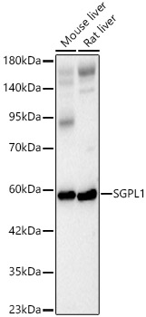
Western blot analysis of various lysates using [KD Validated] SGPL1 Rabbit mAb (A26855) at 1:15000 dilution incubated overnight at 4℃.
Secondary antibody: HRP-conjugated Goat anti-Rabbit IgG (H+L) (AS014) at 1:10000 dilution.
Lysates/proteins: 25 μg per lane.
Blocking buffer: 3% nonfat dry milk in TBST.
Detection: ECL Basic Kit (RM00020).
Exposure time: 45s.
Immunohistochemistry analysis of paraffin-embedded Human kidney tissue using [KD Validated] SGPL1 Rabbit mAb (A26855) at a dilution of 1:200 (40x lens). High pressure antigen retrieval performed with 0.01M Citrate Buffer (pH 6.0) prior to IHC staining.
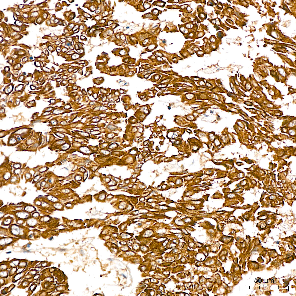
Immunohistochemistry analysis of paraffin-embedded Human liver cancer tissue using [KD Validated] SGPL1 Rabbit mAb (A26855) at a dilution of 1:200 (40x lens). High pressure antigen retrieval performed with 0.01M Citrate Buffer (pH 6.0) prior to IHC staining.
Immunohistochemistry analysis of paraffin-embedded Mouse testis tissue using [KD Validated] SGPL1 Rabbit mAb (A26855) at a dilution of 1:200 (40x lens). High pressure antigen retrieval performed with 0.01M Citrate Buffer (pH 6.0) prior to IHC staining.
Immunohistochemistry analysis of paraffin-embedded Rat testis tissue using [KD Validated] SGPL1 Rabbit mAb (A26855) at a dilution of 1:200 (40x lens). High pressure antigen retrieval performed with 0.01M Citrate Buffer (pH 6.0) prior to IHC staining.
Confocal imaging of paraffin-embedded Mouse small intesine tissue using [KD Validated] SGPL1 Rabbit mAb (A26855, dilution 1:200) followed by a further incubation with Cy3 Goat Anti-Rabbit IgG (H+L) (AS007, dilution 1:500) (Red). DAPI was used for nuclear staining (Blue). High pressure antigen retrieval performed with 0.01M Citrate Buffer(pH 6.0) prior to IF staining. Objective: 40x.
Confocal imaging of paraffin-embedded Rat thymus tissue using [KD Validated] SGPL1 Rabbit mAb (A26855, dilution 1:200) followed by a further incubation with Cy3 Goat Anti-Rabbit IgG (H+L) (AS007, dilution 1:500) (Red). DAPI was used for nuclear staining (Blue). High pressure antigen retrieval performed with 0.01M Citrate Buffer(pH 6.0) prior to IF staining. Objective: 40x.
Confocal imaging of Hep G2 cells using [KD Validated] SGPL1 Rabbit mAb (A26855, dilution 1:200) followed by a further incubation with Cy3 Goat Anti-Rabbit IgG (H+L) (AS007, dilution 1:500) (Red). The cells were counterstained with α-tubulin Mouse mAb (AC012, dilution 1:400) followed by incubation with ABflo® 488-conjugated Goat Anti-Mouse IgG (H+L) Ab (AS076, dilution 1:500) (Green). DAPI was used for nuclear staining (Blue). Objective: 100x.
| Product name | [KD Validated] SGPL1 Rabbit mAb |
|---|---|
| Catalog No. | A26855 |
| Host species | Rabbit |
| Purification method | Affinity purification |
| Isotype | IgG |
| CloneNo. | ARC70997 |
| Immunogen | A synthetic peptide corresponding to a sequence within amino acids 301-400 of human SGPL1 (NP_003892.2). |
|---|---|
| Sequence | VAKLAVKYKIPLHVDACLGGFLIVFMEKAGYPLEHPFDFRVKGVTSISADTHKYGYAPKGSSLVLYSDKKYRNYQFFVDTDWQGGIYASPTIAGSRPGGI |
| Gene ID | |
| Swiss Prot | |
| Synonyms | SPL; S1PL; NPHS14 |
| Calculated MW | 64kDa |
| Observed MW | 60kDa |
| Reactivity | Human, Mouse, Rat |
|---|---|
| Tested applications | WBIHC-PIF/ICCIPChIPChIP-seqRIPFCFC(Intra)ELISAMeDIPNucleotide ArrayDBFACSCoIPCUT&TagmeRIPInhibition |
| Recommended dilution |
|
| Storage buffer | Store at -20℃. Avoid freeze / thaw cycles. Buffer: PBS with 0.09% Sodium azide, 0.05% BSA, 50% glycerol, pH7.3. |
| Key application | Western blotting Immunohistochemistry Immunofluorescence |
| Positive samples | 293T, Hep G2, Rat thymus |
| Cellular location | Endoplasmic reticulum membrane, Single-pass type III membrane protein. |
To download a Certificate of Compliance, please enter your Lot number below:
Lot number
* For research use only. Not for therapeutic or diagnostic purposes.
![ABclonal:Western blot - [KD Validated] SGPL1 Rabbit mAb (A26855)](https://img.abclonal.com/abclonal-manage/Catalog/A26855/A26855_1.jpg?t=1731292302 "ABclonal:Western blot - [KD Validated] SGPL1 Rabbit mAb (A26855)")
![ABclonal:Western blot - [KD Validated] SGPL1 Rabbit mAb (A26855)](https://img.abclonal.com/abclonal-manage/Catalog/A26855/A26855_2.jpg?t=1731292302 "ABclonal:Western blot - [KD Validated] SGPL1 Rabbit mAb (A26855)")
![ABclonal:Immunohistochemistry - [KD Validated] SGPL1 Rabbit mAb (A26855)](https://img.abclonal.com/abclonal-manage/Catalog/A26855/A26855_3.jpg?t=1731292302 "ABclonal:Immunohistochemistry - [KD Validated] SGPL1 Rabbit mAb (A26855)")
![ABclonal:Immunohistochemistry - [KD Validated] SGPL1 Rabbit mAb (A26855)](https://img.abclonal.com/abclonal-manage/Catalog/A26855/A26855_4.jpg?t=1731292302 "ABclonal:Immunohistochemistry - [KD Validated] SGPL1 Rabbit mAb (A26855)")
![ABclonal:Immunohistochemistry - [KD Validated] SGPL1 Rabbit mAb (A26855)](https://img.abclonal.com/abclonal-manage/Catalog/A26855/A26855_5.jpg?t=1731292302 "ABclonal:Immunohistochemistry - [KD Validated] SGPL1 Rabbit mAb (A26855)")
![ABclonal:Immunohistochemistry - [KD Validated] SGPL1 Rabbit mAb (A26855)](https://img.abclonal.com/abclonal-manage/Catalog/A26855/A26855_6.jpg?t=1731292302 "ABclonal:Immunohistochemistry - [KD Validated] SGPL1 Rabbit mAb (A26855)")
![ABclonal:Immunofluorescence - [KD Validated] SGPL1 Rabbit mAb (A26855)](https://img.abclonal.com/abclonal-manage/Catalog/A26855/A26855_7.jpg?t=1731292302 "ABclonal:Immunofluorescence - [KD Validated] SGPL1 Rabbit mAb (A26855)")
![ABclonal:Immunofluorescence - [KD Validated] SGPL1 Rabbit mAb (A26855)](https://img.abclonal.com/abclonal-manage/Catalog/A26855/A26855_8.jpg?t=1731292302 "ABclonal:Immunofluorescence - [KD Validated] SGPL1 Rabbit mAb (A26855)")
![ABclonal:Immunofluorescence - [KD Validated] SGPL1 Rabbit mAb (A26855)](https://img.abclonal.com/abclonal-manage/Catalog/A26855/A26855_9.jpg?t=1731292302 "ABclonal:Immunofluorescence - [KD Validated] SGPL1 Rabbit mAb (A26855)")
![ABclonal:Immunofluorescence - [KD Validated] SGPL1 Rabbit mAb (A26855)](https://img.abclonal.com/abclonal-manage/Catalog/A26855/A26855_10.jpg?t=1731292302 "ABclonal:Immunofluorescence - [KD Validated] SGPL1 Rabbit mAb (A26855)")