Tested applications:WBIHC-PIF/ICCIPChIPChIP-seqRIPFCFC(Intra)ELISAMeDIPNucleotide ArrayDBFACSCoIPCUT&TagmeRIPInhibitionReactivity:Human, Mouse, Rat
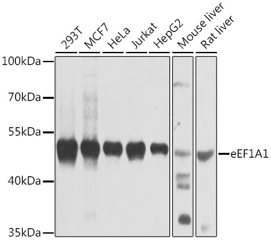
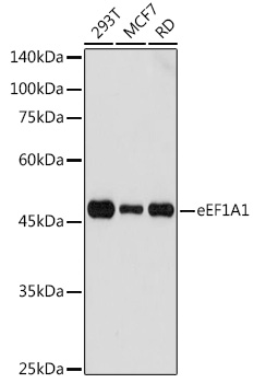
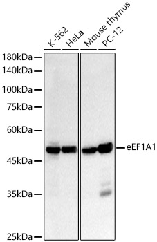
Western blot analysis of various lysates using eEF1A1 Rabbit mAb (A11545) at 1:1000 dilution.
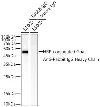
Secondary antibody: HRP-conjugated Goat anti-Rabbit IgG (H+L) (AS014) at 1:10000 dilution.
Lysates/proteins: 25μg per lane.
Blocking buffer: 3% nonfat dry milk in TBST.
Detection: ECL Basic Kit (RM00020).
Exposure time: 90s.
| Product name | eEF1A1 Rabbit mAb |
|---|---|
| Catalog No. | A11545 |
| Host species | Rabbit |
| Purification method | Affinity purification |
| Isotype | IgG |
| CloneNo. | ARC0626 |
| Immunogen | A synthetic peptide corresponding to a sequence within amino acids 250-350 of human eEF1A1 (NP_001393.1). |
|---|---|
| Sequence | LQDVYKIGGIGTVPVGRVETGVLKPGMVVTFAPVNVTTEVKSVEMHHEALSEALPGDNVGFNVKNVSVKDVRRGNVAGDSKNDPPMEAAGFTAQVIILNHP |
| Gene ID | |
| Swiss Prot | |
| Synonyms | CCS3; EF1A; PTI1; CCS-3; EE1A1; EEF-1; EEF1A; EF-Tu; EF1A1; LENG7; eEF1A-1; GRAF-1EF; EF1alpha1; eEF1A1 |
| Calculated MW | 50kDa |
| Observed MW | 50kDa |
| Reactivity | Human, Mouse, Rat |
|---|---|
| Tested applications | WBIHC-PIF/ICCIPChIPChIP-seqRIPFCFC(Intra)ELISAMeDIPNucleotide ArrayDBFACSCoIPCUT&TagmeRIPInhibition |
| Recommended dilution |
|
| Storage buffer | Store at -20℃. Avoid freeze / thaw cycles. Buffer: PBS with 0.05% proclin300, 0.05% BSA, 50% glycerol, pH7.3. |
| Key application | Western blotting Immunohistochemistry Immunofluorescence |
| Positive samples | HeLa, 293T, MCF7, Mouse lung, Mouse liver, Mouse kidney, Rat kidney |
| Cellular location | Cytoplasm, Nucleus, nucleolus. |
| Customer validation | WB(Homo sapiens) |
To download a Certificate of Compliance, please enter your Lot number below:
Lot number
* For research use only. Not for therapeutic or diagnostic purposes.
Publishing research using A11545? Please let us know so that we can cite the reference in this datasheet.
")
")