Tested applications:WBIHC-PIF/ICCIPChIPChIP-seqRIPFCFC(Intra)ELISAMeDIPNucleotide ArrayDBFACSCoIPCUT&TagmeRIPInhibitionReactivity:Human
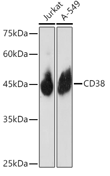
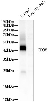
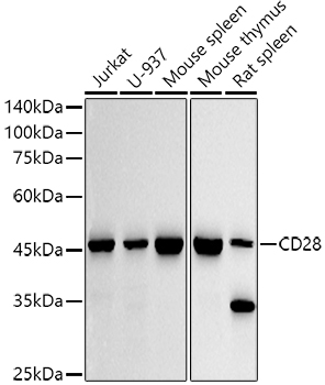
Western blot analysis of various lysates using CD38 Rabbit mAb (A25398) at 1:3000 dilution.
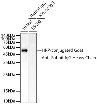
Secondary antibody: HRP-conjugated Goat anti-Rabbit IgG (H+L) (AS014) at 1:10000 dilution.
Lysates/proteins: 25 μg per lane.
Blocking buffer: 3% nonfat dry milk in TBST.
Detection: ECL Basic Kit (RM00020).
Negative control (NC): Hep G2.
Exposure time: 30s.
Immunohistochemistry analysis of paraffin-embedded Human tonsil tissue using CD38 Rabbit mAb (A25398) at a dilution of 1:800 (40x lens). High pressure antigen retrieval was performed with 0.01 M Tris-EDTA buffer (pH 9.0) prior to IHC staining.
Immunohistochemistry analysis of paraffin-embedded Human colon tissue using CD38 Rabbit mAb (A25398) at a dilution of 1:800 (40x lens). High pressure antigen retrieval was performed with 0.01 M Tris-EDTA buffer (pH 9.0) prior to IHC staining.
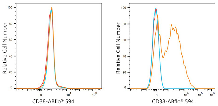
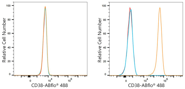
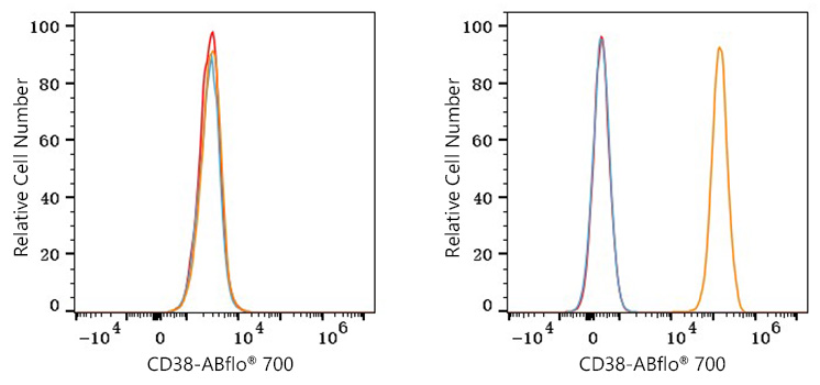
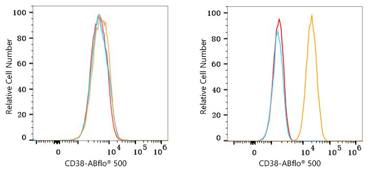
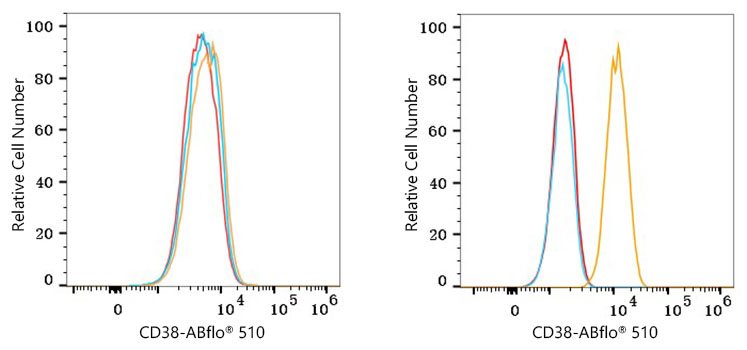
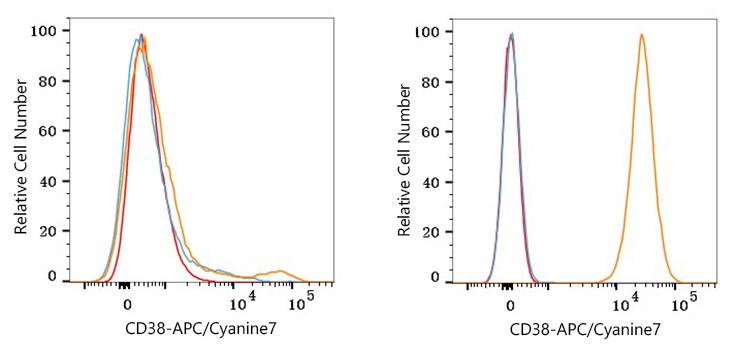
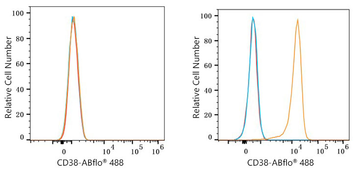
Flow cytometry: 1X10^6 Hep G2 cells (negative control, left) and Daudi cells (right) were surface-stained with CD38 Rabbit mAb (A25398, 2 μg/mL, orange line) or ABflo® 647 Rabbit IgG isotype control (A22070, 5 μl/Test, blue line), followed by Alexa Fluor® 647 conjugated goat anti-rabbit pAb staining. Non-fluorescently stained cells were used as blank control (red line).
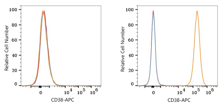
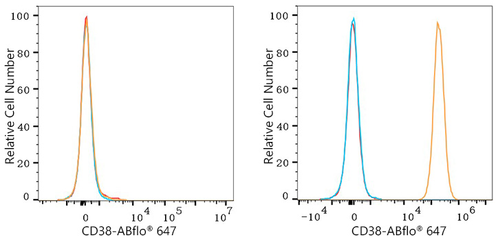
Flow cytometry: 1X10^6 Daudi cells were surface-stained with ABflo® 647 Rabbit IgG isotype control (A22070, 5 μl/Test, left) or CD38 Rabbit mAb (A25398, 2 μg/mL, right).
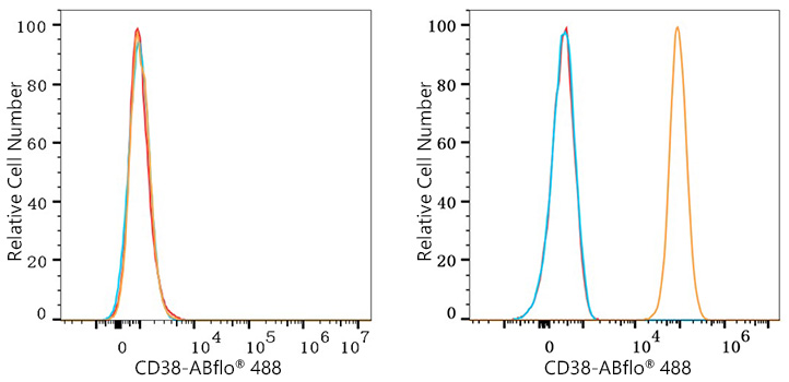
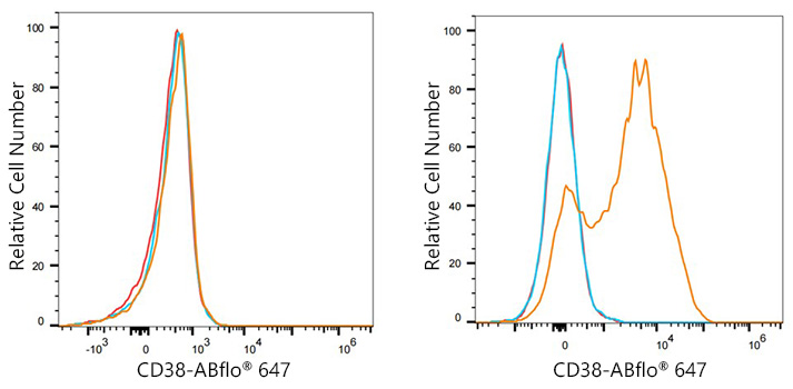
Flow cytometry: 1X10^6 Human PBMC were surface-stained with CD38 Rabbit mAb (A25398, 2 μg/mL, orange line) or ABflo® 647 Rabbit IgG isotype control (A22070, 5 μl/Test, blue line), followed by Alexa Fluor® 647 conjugated goat anti-rabbit pAb staining. Non-fluorescently stained Human PBMC were used as blank control (red line).
| Product name | CD38 Rabbit mAb |
|---|---|
| Catalog No. | A25398 |
| Host species | Rabbit |
| Purification method | Affinity purification |
| Isotype | IgG |
| CloneNo. | ARC66212 |
| Immunogen | Recombinant fusion protein containing a sequence corresponding to amino acids 43-300 of human CD38 (NP_001766.2). |
|---|---|
| Sequence | VPRWRQQWSGPGTTKRFPETVLARCVKYTEIHPEMRHVDCQSVWDAFKGAFISKHPCNITEEDYQPLMKLGTQTVPCNKILLWSRIKDLAHQFTQVQRDMFTLEDTLLGYLADDLTWCGEFNTSKINYQSCPDWRKDCSNNPVSVFWKTVSRRFAEAACDVVHVMLNGSRSKIFDKNSTFGSVEVHNLQPEKVQTLEAWVIHGGREDSRDLCQDPTIKELESIISKRNIQFSCKNIYRPDKFLQCVKNPEDSSCTSEI |
| Gene ID | |
| Swiss Prot | |
| Synonyms | ADPRC1; cADPR1; ADPRC 1 |
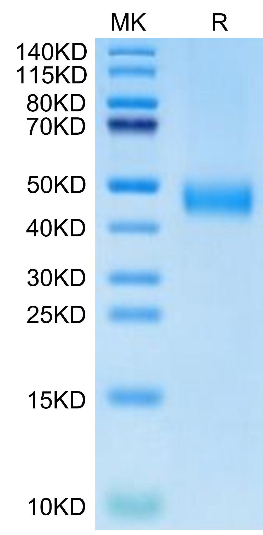
| Calculated MW | 14kDa/34kDa |
| Observed MW | 45kDa |
| Reactivity | Human |
|---|---|
| Tested applications | WBIHC-PIF/ICCIPChIPChIP-seqRIPFCFC(Intra)ELISAMeDIPNucleotide ArrayDBFACSCoIPCUT&TagmeRIPInhibition |
| Recommended dilution |
|
| Storage buffer | Store at -20℃. Avoid freeze / thaw cycles. Buffer: PBS with 0.09% Sodium azide, 0.05% BSA, 50% glycerol, pH7.3. |
| Key application | Western blotting Immunohistochemistry Flow Cytometry |
| Positive samples | Daudi, Ramos |
| Cellular location | Membrane, Single-pass type II membrane protein, basolateral plasma membrane, cell surface, extracellular exosome, nucleus, plasma membrane. |
To download a Certificate of Compliance, please enter your Lot number below:
Lot number
* For research use only. Not for therapeutic or diagnostic purposes.
")
")
")
")
")
")
")