Tested applications:WBIHC-PIF/ICCIPChIPChIP-seqRIPFCFC(Intra)ELISAMeDIPNucleotide ArrayDBFACSCoIPCUT&TagmeRIPInhibitionReactivity:Human, Mouse, Rat, Monkey
Western blot analysis of lysates from wild type (WT) and eIF4E knockdown (KD) 293T cells using [KD Validated] eIF4E Rabbit mAb (A25608) at 1:3000 dilution.
Secondary antibody: HRP-conjugated Goat anti-Rabbit IgG (H+L) (AS014) at 1:10000 dilution.
Lysates/proteins: 25 μg per lane.
Blocking buffer: 3% nonfat dry milk in TBST.
Detection: ECL Basic Kit (RM00020).
Exposure time: 1s.
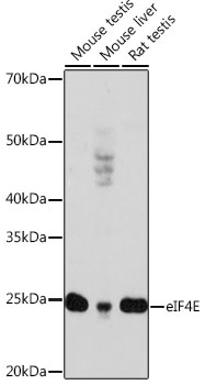
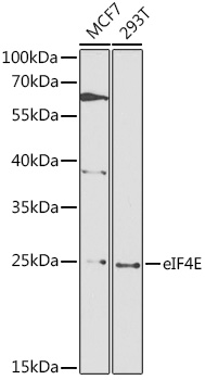
Western blot analysis of various lysates using [KD Validated] eIF4E Rabbit mAb (A25608) at 1:3000 dilution.
Secondary antibody: HRP-conjugated Goat anti-Rabbit IgG (H+L) (AS014) at 1:10000 dilution.
Lysates/proteins: 25 μg per lane.
Blocking buffer: 3% nonfat dry milk in TBST.
Detection: ECL Basic Kit (RM00020).
Exposure time: 10s.
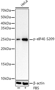
Western blot analysis of various lysates using [KD Validated] eIF4E Rabbit mAb (A25608) at 1:3000 dilution incubated overnight at 4℃.
Secondary antibody: HRP-conjugated Goat anti-Rabbit IgG (H+L) (AS014) at 1:10000 dilution.
Lysates/proteins: 25 μg per lane.
Blocking buffer: 3% nonfat dry milk in TBST.
Detection: ECL Basic Kit (RM00020).
Exposure time: 10s.
Confocal imaging of 293T cells using [KD Validated] eIF4E Rabbit mAb (A25608, dilution 1:200) followed by a further incubation with Cy3 Goat Anti-Rabbit IgG (H+L) (AS007, dilution 1:500) (Red). DAPI was used for nuclear staining (Blue). Objective: 100x.
Confocal imaging of Hep G2 cells using [KD Validated] eIF4E Rabbit mAb (A25608, dilution 1:200) followed by a further incubation with Cy3 Goat Anti-Rabbit IgG (H+L) (AS007, dilution 1:500) (Red). The cells were counterstained with α-Tubulin Mouse mAb (AC012, dilution 1:400) followed by incubation with ABflo® 488-conjugated Goat Anti-Mouse IgG (H+L) Ab (AS076, dilution 1:500) (Green). DAPI was used for nuclear staining (Blue). Objective: 100x.
Confocal imaging of NIH/3T3 cells using [KD Validated] eIF4E Rabbit mAb (A25608, dilution 1:200) followed by a further incubation with Cy3 Goat Anti-Rabbit IgG (H+L) (AS007, dilution 1:500) (Red). The cells were counterstained with α-Tubulin Mouse mAb (AC012, dilution 1:400) followed by incubation with ABflo® 488-conjugated Goat Anti-Mouse IgG (H+L) Ab (AS076, dilution 1:500) (Green). DAPI was used for nuclear staining (Blue). Objective: 100x.
Confocal imaging of PC-12 cells using [KD Validated] eIF4E Rabbit mAb (A25608, dilution 1:200) followed by a further incubation with Cy3 Goat Anti-Rabbit IgG (H+L) (AS007, dilution 1:500) (Red). The cells were counterstained with α-Tubulin Mouse mAb (AC012, dilution 1:400) followed by incubation with ABflo® 488-conjugated Goat Anti-Mouse IgG (H+L) Ab (AS076, dilution 1:500) (Green). DAPI was used for nuclear staining (Blue). Objective: 100x.
| Product name | [KD Validated] eIF4E Rabbit mAb |
|---|---|
| Catalog No. | A25608 |
| Host species | Rabbit |
| Purification method | Affinity purification |
| Isotype | IgG |
| CloneNo. | ARC66295 |
| Immunogen | A synthetic peptide corresponding to a sequence within amino acids 1-100 of human eIF4E (NP_001959.1). |
|---|---|
| Sequence | MATVEPETTPTPNPPTTEEEKTESNQEVANPEHYIKHPLQNRWALWFFKNDKSKTWQANLRLISKFDTVEDFWALYNHIQLSSNLMPGCDYSLFKDGIEP |
| Gene ID | |
| Swiss Prot | |
| Synonyms | CBP; EIF4F; AUTS19; EIF4E1; eIF-4E; EIF4EL1 |
| Calculated MW | 25kDa/27kDa/29kDa |
| Observed MW | 28kDa/ |
| Reactivity | Human, Mouse, Rat, Monkey |
|---|---|
| Tested applications | WBIHC-PIF/ICCIPChIPChIP-seqRIPFCFC(Intra)ELISAMeDIPNucleotide ArrayDBFACSCoIPCUT&TagmeRIPInhibition |
| Recommended dilution |
|
| Storage buffer | Store at -20℃. Avoid freeze / thaw cycles. Buffer: PBS with 0.05% proclin300, 0.05% BSA, 50% glycerol, pH7.3. |
| Key application | Western blotting Immunofluorescence Immunoprecipitation |
| Positive samples | 293T, HeLa, COS-7, NIH/3T3, RAW 264.7 |
| Cellular location | Cytoplasm, P-body, Stress granule, Cytosol, Nucleus, P-body, chromatoid body, cytoplasm, cytoplasmic ribonucleoprotein granule, cytoplasmic stress granule, cytosol, eukaryotic translation initiation factor 4F complex, extracellular exosome, glutamatergic synapse, nucleus, perinuclear region of cytoplasm, postsynaptic cytosol. |
To download a Certificate of Compliance, please enter your Lot number below:
Lot number
* For research use only. Not for therapeutic or diagnostic purposes.
![ABclonal:Western blot - [KD Validated] eIF4E Rabbit mAb (A25608)](https://img.abclonal.com/abclonal-manage/Catalog/A25608/A25608_1.jpg?t=1737682644 "ABclonal:Western blot - [KD Validated] eIF4E Rabbit mAb (A25608)")
![ABclonal:Western blot - [KD Validated] eIF4E Rabbit mAb (A25608)](https://img.abclonal.com/abclonal-manage/Catalog/A25608/A25608_2.jpg?t=1737682644 "ABclonal:Western blot - [KD Validated] eIF4E Rabbit mAb (A25608)")
![ABclonal:Western blot - [KD Validated] eIF4E Rabbit mAb (A25608)](https://img.abclonal.com/abclonal-manage/Catalog/A25608/A25608_3.jpg?t=1737682644 "ABclonal:Western blot - [KD Validated] eIF4E Rabbit mAb (A25608)")
![ABclonal:Immunofluorescence - [KD Validated] eIF4E Rabbit mAb (A25608)](https://img.abclonal.com/abclonal-manage/Catalog/A25608/A25608_4.jpg?t=1737682644 "ABclonal:Immunofluorescence - [KD Validated] eIF4E Rabbit mAb (A25608)")
![ABclonal:Immunofluorescence - [KD Validated] eIF4E Rabbit mAb (A25608)](https://img.abclonal.com/abclonal-manage/Catalog/A25608/A25608_5.jpg?t=1737682644 "ABclonal:Immunofluorescence - [KD Validated] eIF4E Rabbit mAb (A25608)")
![ABclonal:Immunofluorescence - [KD Validated] eIF4E Rabbit mAb (A25608)](https://img.abclonal.com/abclonal-manage/Catalog/A25608/A25608_6.jpg?t=1737682644 "ABclonal:Immunofluorescence - [KD Validated] eIF4E Rabbit mAb (A25608)")
![ABclonal:Immunofluorescence - [KD Validated] eIF4E Rabbit mAb (A25608)](https://img.abclonal.com/abclonal-manage/Catalog/A25608/A25608_7.jpg?t=1737682644 "ABclonal:Immunofluorescence - [KD Validated] eIF4E Rabbit mAb (A25608)")
![ABclonal:Immunoprecipitation - [KD Validated] eIF4E Rabbit mAb (A25608)](https://img.abclonal.com/abclonal-manage/Catalog/A25608/A25608_8.jpg?t=1737682644 "ABclonal:Immunoprecipitation - [KD Validated] eIF4E Rabbit mAb (A25608)")
![[KD Validated] eIF4E Rabbit pAb](https://img.abclonal.com/abclonal-manage/Catalog/A2162/A2162_1.jpg?t=1728975908)
_WB_01.jpg?t=1717400688)