Tested applications:WBIHC-PIF/ICCIPChIPChIP-seqRIPFCFC(Intra)ELISAMeDIPNucleotide ArrayDBFACSCoIPCUT&TagmeRIPInhibitionReactivity:Human, Mouse, Rat, Other (Wide Range Predicted)
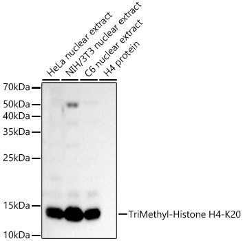
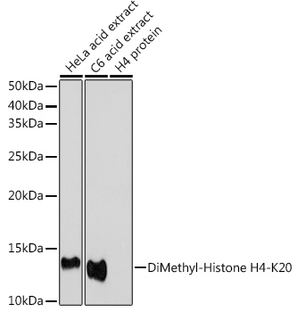
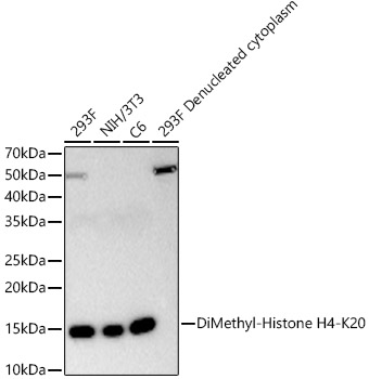
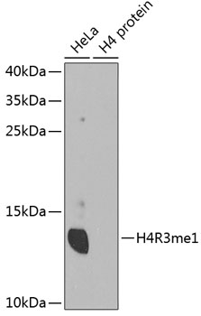
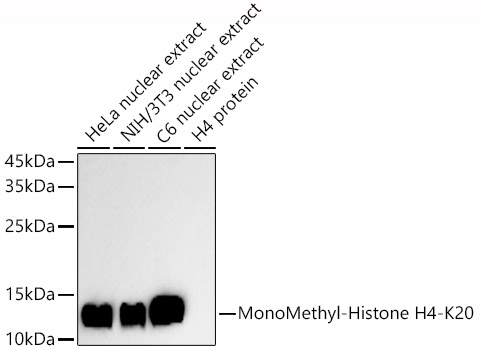
Western blot analysis of various lysates using TriMethyl-Histone H4-K20 Rabbit mAb (A27268) at 1:16000 dilution incubated overnight at 4℃.
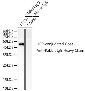
Secondary antibody: HRP-conjugated Goat anti-Rabbit IgG (H+L) (AS014) at 1:10000 dilution.
Lysates/proteins: 25 μg per lane.
Blocking buffer: 3% nonfat dry milk in TBST.
Detection: ECL Basic Kit (RM00020).
Negative control (NC): H4 protein
Exposure time: 1s.
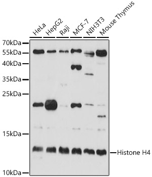
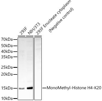
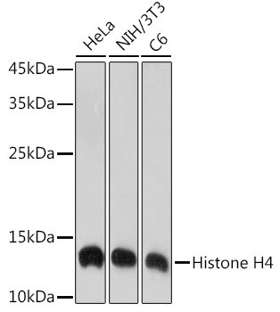
Western blot analysis of various lysates using TriMethyl-Histone H4-K20 Rabbit mAb (A27268) at 1:16000 dilution incubated overnight at 4℃.
Secondary antibody: HRP-conjugated Goat anti-Rabbit IgG (H+L) (AS014) at 1:10000 dilution.
Lysates/proteins: 25 μg per lane.
Blocking buffer: 3% nonfat dry milk in TBST.
Detection: ECL Basic Kit (RM00020).
Negative control (NC): H4 protein
Exposure time: 20s.
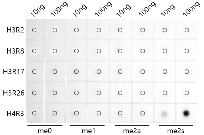
Dot-blot analysis of all sorts of peptides using TriMethyl-Histone H4-K20 Rabbit mAb (A27268) at 1:1000 dilution.
Immunohistochemistry analysis of paraffin-embedded Mouse lung tissue using TriMethyl-Histone H4-K20 Rabbit mAb (A27268) at a dilution of 1:500 (40x lens). High pressure antigen retrieval performed with 0.01M Citrate Buffer (pH 6.0) prior to IHC staining.
Immunohistochemistry analysis of paraffin-embedded Human thyroid cancer tissue using TriMethyl-Histone H4-K20 Rabbit mAb (A27268) at a dilution of 1:500 (40x lens). High pressure antigen retrieval performed with 0.01M Citrate Buffer (pH 6.0) prior to IHC staining.
Immunohistochemistry analysis of paraffin-embedded Rat colon tissue using TriMethyl-Histone H4-K20 Rabbit mAb (A27268) at a dilution of 1:500 (40x lens). High pressure antigen retrieval performed with 0.01M Citrate Buffer (pH 6.0) prior to IHC staining.
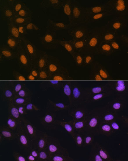
Confocal imaging of NIH/3T3 cells using TriMethyl-Histone H4-K20 Rabbit mAb (A27268, dilution 1:200) followed by a further incubation with Cy3 Goat Anti-Rabbit IgG (H+L) (AS007, dilution 1:500) (Red). The cells were counterstained with α-Tubulin Mouse mAb (AC012, dilution 1:400) followed by incubation with ABflo® 488-conjugated Goat Anti-Mouse IgG (H+L) Ab (AS076, dilution 1:500) (Green). DAPI was used for nuclear staining (Blue). Objective: 100x.
Confocal imaging of HeLa cells using TriMethyl-Histone H4-K20 Rabbit mAb (A27268, dilution 1:200) followed by a further incubation with Cy3 Goat Anti-Rabbit IgG (H+L) (AS007, dilution 1:500) (Red). The cells were counterstained with α-Tubulin Mouse mAb (AC012, dilution 1:400) followed by incubation with ABflo® 488-conjugated Goat Anti-Mouse IgG (H+L) Ab (AS076, dilution 1:500) (Green). DAPI was used for nuclear staining (Blue). Objective: 100x.
| Product name | TriMethyl-Histone H4-K20 Rabbit mAb |
|---|---|
| Catalog No. | A27268 |
| Host species | Rabbit |
| Purification method | Affinity purification |
| Isotype | IgG |
| CloneNo. | ARC66120 |
| Immunogen | A synthetic trimethylated peptide around K20 of human Histone H4 (NP_003539.1). |
|---|---|
| Sequence | HRKVL |
| Gene ID | |
| Swiss Prot | |
| Synonyms | H4; H4/n; H4C1; H4C2; H4C3; H4C4; H4C5; H4C6; H4C8; H4C9; H4F2; H4FN; FO108; H4-16; H4C11; H4C12; H4C13; H4C15; H4C16; HIST2H4; HIST2H4A |
| Calculated MW | 11kDa |
| Observed MW | 11kDa |
| Reactivity | Human, Mouse, Rat, Other (Wide Range Predicted) |
|---|---|
| Tested applications | WBIHC-PIF/ICCIPChIPChIP-seqRIPFCFC(Intra)ELISAMeDIPNucleotide ArrayDBFACSCoIPCUT&TagmeRIPInhibition |
| Recommended dilution |
|
| Storage buffer | Store at -20℃. Avoid freeze / thaw cycles. Buffer: PBS with 0.09% Sodium azide, 0.05% BSA, 50% glycerol, pH7.3. |
| Key application | Western blotting Immunohistochemistry Immunofluorescence |
| Positive samples | HeLa, C6, NIH/3T3 |
| Cellular location | Chromosome, Nucleus. |
To download a Certificate of Compliance, please enter your Lot number below:
Lot number
* For research use only. Not for therapeutic or diagnostic purposes.
")
")
")
")
")
")
")
")
_WB_01.jpg?t=1721361071)
_WB_01.jpg?t=1715822831)
_WB_01.jpg?t=1715651707)
_WB_01.jpg?t=1713322500)
_WB_01.jpg?t=1715675057)
_WB_01.jpg?t=1721355659)
_WB_01.jpg?t=1718355783)

_WB_01.jpg?t=1716191102)